-
Reagents
- Flow Cytometry Reagents
-
Western Blotting and Molecular Reagents
- Immunoassay Reagents
-
Single-Cell Multiomics Reagents
- BD® OMICS-Guard Sample Preservation Buffer
- BD® AbSeq Assay
- BD® OMICS-One Immune Profiler Protein Panel
- BD® Single-Cell Multiplexing Kit
- BD Rhapsody™ ATAC-Seq Assays
- BD Rhapsody™ Whole Transcriptome Analysis (WTA) Amplification Kit
- BD Rhapsody™ TCR/BCR Next Multiomic Assays
- BD Rhapsody™ Targeted mRNA Kits
- BD Rhapsody™ Accessory Kits
-
Functional Assays
-
Microscopy and Imaging Reagents
-
Cell Preparation and Separation Reagents
-
- BD® OMICS-Guard Sample Preservation Buffer
- BD® AbSeq Assay
- BD® OMICS-One Immune Profiler Protein Panel
- BD® Single-Cell Multiplexing Kit
- BD Rhapsody™ ATAC-Seq Assays
- BD Rhapsody™ Whole Transcriptome Analysis (WTA) Amplification Kit
- BD Rhapsody™ TCR/BCR Next Multiomic Assays
- BD Rhapsody™ Targeted mRNA Kits
- BD Rhapsody™ Accessory Kits
- United States (English)
-
Change country/language
Old Browser
This page has been recently translated and is available in French now.
Looks like you're visiting us from {countryName}.
Would you like to stay on the current country site or be switched to your country?


Regulatory Status Legend
Any use of products other than the permitted use without the express written authorization of Becton, Dickinson and Company is strictly prohibited.
Preparation And Storage
Recommended Assay Procedures
BD® CompBeads can be used as surrogates to assess fluorescence spillover (compensation). When fluorochrome conjugated antibodies are bound to BD® CompBeads, they have spectral properties very similar to cells. However, for some fluorochromes there can be small differences in spectral emissions compared to cells, resulting in spillover values that differ when compared to biological controls. It is strongly recommended that when using a reagent for the first time, users compare the spillover on cells and BD® CompBeads to ensure that BD® CompBeads are appropriate for your specific cellular application.
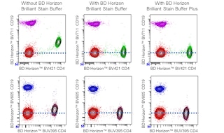
For optimal and reproducible results, BD Horizon Brilliant Stain Buffer should be used anytime BD Horizon Brilliant dyes are used in a multicolor flow cytometry panel. Fluorescent dye interactions may cause staining artifacts which may affect data interpretation. The BD Horizon Brilliant Stain Buffer was designed to minimize these interactions. When BD Horizon Brilliant Stain Buffer is used in in the multicolor panel, it should also be used in the corresponding compensation controls for all dyes to achieve the most accurate compensation. For the most accurate compensation, compensation controls created with either cells or beads should be exposed to BD Horizon Brilliant Stain Buffer for the same length of time as the corresponding multicolor panel. More information can be found in the Technical Data Sheet of the BD Horizon Brilliant Stain Buffer (Cat. No. 563794/566349) or the BD Horizon Brilliant Stain Buffer Plus (Cat. No. 566385).
Product Notices
- The production process underwent stringent testing and validation to assure that it generates a high-quality conjugate with consistent performance and specific binding activity. However, verification testing has not been performed on all conjugate lots.
- Researchers should determine the optimal concentration of this reagent for their individual applications.
- An isotype control should be used at the same concentration as the antibody of interest.
- Caution: Sodium azide yields highly toxic hydrazoic acid under acidic conditions. Dilute azide compounds in running water before discarding to avoid accumulation of potentially explosive deposits in plumbing.
- For fluorochrome spectra and suitable instrument settings, please refer to our Multicolor Flow Cytometry web page at www.bdbiosciences.com/colors.
- Please refer to www.bdbiosciences.com/us/s/resources for technical protocols.
- BD Horizon Brilliant Stain Buffer is covered by one or more of the following US patents: 8,110,673; 8,158,444; 8,575,303; 8,354,239.
- Please refer to http://regdocs.bd.com to access safety data sheets (SDS).
- BD Horizon Brilliant™ Violet 750 is covered by one or more of the following US patents: 8,158,444; 8,802,450; 8,575,303; 8,455,613; 8,227,187; 8,841,072; 8,110,673.
- Human donor specific background has been observed in relation to the presence of anti-polyethylene glycol (PEG) antibodies, developed as a result of certain vaccines containing PEG, including some COVID-19 vaccines. We recommend use of BD Horizon Brilliant™ Stain Buffer in your experiments to help mitigate potential background. For more information visit https://www.bdbiosciences.com/en-us/support/product-notices.
Companion Products


The TS2/16 monoclonal antibody specifically recognizes CD29 which is also known as Integrin beta-1 (Integrin β1 or β1 Integrin), Glycoprotein IIa (GPIIA), Very late activation protein beta (VLA-beta; VLAB), and Fibronectin receptor subunit beta (FNRB). CD29 is an ~130 kDa single pass type I transmembrane glycoprotein that is encoded by ITGB1 (Integrin subunit beta 1). β1 integrins constitute the largest subgroup of integrins as they form noncovalent heterodimeric complexes with at least 12 different alpha integrin (Integrin α) subunits. These heterodimeric β1 integrins mediate a variety of interactions between cells and between cells and the extracellular matrix that are involved in cellular signaling, growth, survival, adhesion, and migration. The family of heterodimeric β1 integrins includes receptors for vascular cell adhesion molecule 1 (VCAM-1), extracellular matrix (ECM) components such as collagen, fibronectin, laminin, and vitronectin, and some microbial ligands. The β1 integrin subunit is widely expressed on hematopoietic and non-hematopoietic cells including T and B cells, dendritic cells (DC), NK cells, monocytes and macrophages, granulocytes, as well as mast cells, fibroblasts, endothelial cells, epithelial cells, and stem cells. Platelets express high levels of CD29 (β1 integrin) and may be bound to leucocytes from human blood. Gating out cells expressing the platelet marker CD41a will avoid attributing platelet CD29 (β1 integrin) expression to attached leucocytes. The TS2/16 antibody is reportedly useful for multiple applications including flow cytometry, immunohistochemistry, and functional studies.

Development References (12)
-
Hemler ME, Bodorova J, Kawaguchi S, Weitzman J, Kassner P. Adhesion structures subpanel 6, β1 integrins/VLA: CD29/CD49. In: Schlossman SF. Stuart F. Schlossman .. et al., ed. Leucocyte typing V : white cell differentiation antigens : proceedings of the fifth international workshop and conference held in Boston, USA, 3-7 November, 1993. Oxford: Oxford University Press; 1995:1609-1612.
-
Hemler ME, Bodorova J, Pasqualini R. CD29 (integrin β1) cluster report. In: Schlossman SF. Stuart F. Schlossman .. et al., ed. Leucocyte typing V : white cell differentiation antigens : proceedings of the fifth international workshop and conference held in Boston, USA, 3-7 November, 1993. Oxford: Oxford University Press; 1995:1612-1613.
-
Hemler ME, Sanchez-Madrid F, Flotte TJ, et al. Glycoproteins of 210,000 and 130,000 m.w. on activated T cells: cell distribution and antigenic relation to components on resting cells and T cell lines.. J Immunol. 1984; 132(6):3011-8. (Immunogen: Immunohistochemistry, Immunoprecipitation, Radioimmunoassay). View Reference
-
Logdberg L, West S, Vattay A, Dottavio D. Soluble recombinant integrin β1 (CD29) as a probe to identify CD29-reactive mAb. In: Schlossman SF. Stuart F. Schlossman .. et al., ed. Leucocyte typing V : white cell differentiation antigens : proceedings of the fifth international workshop and conference held in Boston, USA, 3-7 November, 1993. Oxford: Oxford University Press; 1995:1626-1629.
-
Luque A, Gomez M, Puzon W, Takada Y, Sanchez-Madrid F, Cabanas C. Activated conformations of very late activation integrins detected by a group of antibodies (HUTS) specific for a novel regulatory region (355-425) of the common beta 1 chain. J Biol Chem. 1996; 271(19):11067-11075. (Clone-specific: Flow cytometry, Functional assay, Immunoaffinity chromatography). View Reference
-
Luque A, Sánchez-Madrid F, Cabañas C. Functional regulation of the human integrin VLA-1 (CD49a/CD29) by divalent cations and stimulatory beta 1 antibodies.. FEBS Lett. 1994; 346(2-3):278-84. (Clone-specific: Flow cytometry). View Reference
-
Masumoto A, Hemler ME. Mutation of putative divalent cation sites in the alpha 4 subunit of the integrin VLA-4: distinct effects on adhesion to CS1/fibronectin, VCAM-1, and invasin.. J Cell Biol. 1993; 123(1):245-53. (Clone-specific: Functional assay). View Reference
-
Mould AP, Garratt AN, Puzon-McLaughlin W, Takada Y, Humphries MJ. Regulation of integrin function: evidence that bivalent-cation-induced conformational changes lead to the unmasking of ligand-binding sites within integrin alpha5 beta1.. Biochem J. 1998; 331 ( Pt 3):821-8. (Clone-specific: Blocking, ELISA). View Reference
-
Springer TA, Luther E, Klickstein LB. Adhesion structures: section report. In: Schlossman SF. Schlossman SF, Boumsell L, Gilks W, et al, ed. Leucocyte Typing V: White Cell Differentiation Antigens. New York, NY: Oxford; 1995:1443-1467.
-
Tsuchida J, Ueki S, Saito Y, Takagi J. Classification of 'activation' antibodies against integrin beta1 chain.. FEBS Lett. 1997; 416(2):212-6. (Clone-specific: Activation, Functional assay, Radioimmunoassay). View Reference
-
Walsh GM, Symon FA, Lazarovils AL, Wardlaw AJ. Integrin alpha 4 beta 7 mediates human eosinophil interaction with MAdCAM-1, VCAM-1 and fibronectin.. Immunology. 1996; 89(1):112-9. (Clone-specific: Functional assay). View Reference
-
Zutter MM. Immunohistochemistry of Adhesion Structure Subpanel 6 mAb to β1 (CD29/CD49) integrins. In: Schlossman SF. Stuart F. Schlossman .. et al., ed. Leucocyte typing V : white cell differentiation antigens : proceedings of the fifth international workshop and conference held in Boston, USA, 3-7 November, 1993. Oxford: Oxford University Press; 1995:1621-1623.
Please refer to Support Documents for Quality Certificates
Global - Refer to manufacturer's instructions for use and related User Manuals and Technical data sheets before using this products as described
Comparisons, where applicable, are made against older BD Technology, manual methods or are general performance claims. Comparisons are not made against non-BD technologies, unless otherwise noted.
For Research Use Only. Not for use in diagnostic or therapeutic procedures.