-
Reagents
- Flow Cytometry Reagents
-
Western Blotting and Molecular Reagents
- Immunoassay Reagents
-
Single-Cell Multiomics Reagents
- BD® OMICS-Guard Sample Preservation Buffer
- BD® AbSeq Assay
- BD® OMICS-One Immune Profiler Protein Panel
- BD® Single-Cell Multiplexing Kit
- BD Rhapsody™ ATAC-Seq Assays
- BD Rhapsody™ Whole Transcriptome Analysis (WTA) Amplification Kit
- BD Rhapsody™ TCR/BCR Next Multiomic Assays
- BD Rhapsody™ Targeted mRNA Kits
- BD Rhapsody™ Accessory Kits
-
Functional Assays
-
Microscopy and Imaging Reagents
-
Cell Preparation and Separation Reagents
-
- BD® OMICS-Guard Sample Preservation Buffer
- BD® AbSeq Assay
- BD® OMICS-One Immune Profiler Protein Panel
- BD® Single-Cell Multiplexing Kit
- BD Rhapsody™ ATAC-Seq Assays
- BD Rhapsody™ Whole Transcriptome Analysis (WTA) Amplification Kit
- BD Rhapsody™ TCR/BCR Next Multiomic Assays
- BD Rhapsody™ Targeted mRNA Kits
- BD Rhapsody™ Accessory Kits
- United States (English)
-
Change country/language
Old Browser
This page has been recently translated and is available in French now.
Looks like you're visiting us from {countryName}.
Would you like to stay on the current country site or be switched to your country?


Regulatory Status Legend
Any use of products other than the permitted use without the express written authorization of Becton, Dickinson and Company is strictly prohibited.
Preparation And Storage
Recommended Assay Procedures
For optimal and reproducible results, BD Horizon Brilliant Stain Buffer should be used anytime two or more BD Horizon Brilliant dyes (including BD OptiBuild Brilliant reagents) are used in the same experiment. Fluorescent dye interactions may cause staining artifacts which may affect data interpretation. The BD Horizon Brilliant Stain Buffer was designed to minimize these interactions. More information can be found in the Technical Data Sheet of the BD Horizon Brilliant Stain Buffer (Cat. No. 563794).
Product Notices
- This antibody was developed for use in flow cytometry.
- The production process underwent stringent testing and validation to assure that it generates a high-quality conjugate with consistent performance and specific binding activity. However, verification testing has not been performed on all conjugate lots.
- Researchers should determine the optimal concentration of this reagent for their individual applications.
- An isotype control should be used at the same concentration as the antibody of interest.
- Caution: Sodium azide yields highly toxic hydrazoic acid under acidic conditions. Dilute azide compounds in running water before discarding to avoid accumulation of potentially explosive deposits in plumbing.
- For fluorochrome spectra and suitable instrument settings, please refer to our Multicolor Flow Cytometry web page at www.bdbiosciences.com/colors.
- Please refer to www.bdbiosciences.com/us/s/resources for technical protocols.
- BD Horizon Brilliant Stain Buffer is covered by one or more of the following US patents: 8,110,673; 8,158,444; 8,575,303; 8,354,239.
- BD Horizon Brilliant Violet 510 is covered by one or more of the following US patents: 8,575,303; 8,354,239.
Companion Products


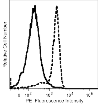
The 11F2 monoclonal antibody specifically reacts with a framework epitope of the γ/δ TCR. The γ/δ TCR is a heterodimeric glycoprotein that is noncovalently associated with the CD3 antigen complex. The γ and δ TCR chains are composed of constant and variable regions, each encoded by distinct gene segments. The γ chain forms either disulfide-linked or non-disulfide-linked heterodimers with the δ-subunit. TCR γ/δ is present on a minor subset of T lymphocytes in peripheral blood, thymus, spleen, and lymph node. TCR γ/δ-positive T lymphocytes comprise 1% to 9% of normal peripheral blood lymphocytes and less than 2% of normal thymocytes. The 11F2 antibody is mitogenic for γ/δ-TCR-bearing T lymphocytes.
The antibody was conjugated to BD Horizon™ BV510 which is part of the BD Horizon Brilliant™ Violet family of dyes. With an Ex Max of 405-nm and Em Max at 510-nm, BD Horizon BV510 can be excited by the violet laser and detected in the BD Horizon V500 (525/50-nm) filter set. BD Horizon BV510 conjugates are useful for the detection of dim markers off the violet laser.

Development References (15)
-
Blink SE, Miller SD. The contribution of gammadelta T cells to the pathogenesis of EAE and MS.. Curr Mol Med. 2009; 9(1):15-22. (Biology). View Reference
-
Bonneville M, O'Brien RL, Born WK. Gammadelta T cell effector functions: a blend of innate programming and acquired plasticity. Nat Rev Immunol. 2110; 10(7):467-478. (Biology). View Reference
-
Borst J, van Dongen JJ, Bolhuis RL, et al. Distinct molecular forms of human T cell receptor gamma/delta detected on viable T cells by a monoclonal antibody.. J Exp Med. 1988; 167(5):1625-44. (Immunogen: Electron microscopy, ELISA, Flow cytometry, Fluorescence microscopy, Immunofluorescence). View Reference
-
Cairo C, Hebbeler AM, Propp N, Bryant JL, Colizzi V, Pauza CD. Innate-like gammadelta T cell responses to mycobacterium Bacille Calmette-Guerin using the public V gamma 2 repertoire in Macaca fascicularis.. Tuberculosis (Edinb). 2007; 87(4):373-83. (Biology). View Reference
-
Carding SR, Egan PJ. The importance of gamma delta T cells in the resolution of pathogen-induced inflammatory immune responses.. Immunol Rev. 2000; 173:98-108. (Biology). View Reference
-
Chen ZW. Immune regulation of gammadelta T cell responses in mycobacterial infections.. Clin Immunol. 2005; 116(3):202-7. (Biology). View Reference
-
García VE, Sieling PA, Gong J, et al. Single-cell cytokine analysis of gamma delta T cell responses to nonpeptide mycobacterial antigens.. J Immunol. 1997; 159(3):1328-35. (Biology). View Reference
-
Huang D, Chen CY, Zhang M, et al. Clonal immune responses of Mycobacterium-specific γδ T cells in tuberculous and non-tuberculous tissues during M. tuberculosis infection.. PLoS ONE. 2012; 7(2):e30631. (Biology). View Reference
-
Ichinohasama R, Miura I, Takahashi T, et al. Peripheral CD4+ CD8- gammadelta T cell lymphoma: a case report with multiparameter analyses.. Hum Pathol. 1996; 27(12):1370-7. (Clone-specific). View Reference
-
Lanier LL, Federspiel NA, Ruitenberg JJ, et al. The T cell antigen receptor complex expressed on normal peripheral blood CD4-, CD8- T lymphocytes. A CD3-associated disulfide-linked gamma chain heterodimer.. J Exp Med. 1987; 165(4):1076-94. (Biology). View Reference
-
Lanier LL, Ruitenberg J, Bolhuis RL, Borst J, Phillips JH, Testi R. Structural and serological heterogeneity of gamma/delta T cell antigen receptor expression in thymus and peripheral blood.. Eur J Immunol. 1988; 18(12):1985-92. (Biology). View Reference
-
Lanier LL, Serafini AT, Ruitenberg JJ, et al. The gamma T-cell antigen receptor.. J Clin Immunol. 1987; 7(6):429-40. (Biology). View Reference
-
Testi R, Lanier LL. Functional expression of CD28 on T cell antigen receptor gamma/delta-bearing T lymphocytes.. Eur J Immunol. 1989; 19(1):185-8. (Biology). View Reference
-
Urban EM, Chapoval AI, Pauza CD. Repertoire development and the control of cytotoxic/effector function in human gammadelta T cells.. Clin Dev Immunol. 2010; 2010:732893. (Biology). View Reference
-
Voogt PJ, Falkenburg JH, Fibbe WE, et al. Normal hematopoietic progenitor cells and malignant lymphohematopoietic cells show different susceptibility to direct cell-mediated MHC-non-restricted lysis by T cell receptor-/CD3-, T cell receptor gamma delta+/CD3+ and T cell receptor-alpha beta+/CD3+ lymphocytes.. J Immunol. 1989; 142(5):1774-80. (Biology). View Reference
Please refer to Support Documents for Quality Certificates
Global - Refer to manufacturer's instructions for use and related User Manuals and Technical data sheets before using this products as described
Comparisons, where applicable, are made against older BD Technology, manual methods or are general performance claims. Comparisons are not made against non-BD technologies, unless otherwise noted.
For Research Use Only. Not for use in diagnostic or therapeutic procedures.