-
Reagents
- Flow Cytometry Reagents
-
Western Blotting and Molecular Reagents
- Immunoassay Reagents
-
Single-Cell Multiomics Reagents
- BD® OMICS-Guard Sample Preservation Buffer
- BD® AbSeq Assay
- BD® OMICS-One Immune Profiler Protein Panel
- BD® Single-Cell Multiplexing Kit
- BD Rhapsody™ ATAC-Seq Assays
- BD Rhapsody™ Whole Transcriptome Analysis (WTA) Amplification Kit
- BD Rhapsody™ TCR/BCR Next Multiomic Assays
- BD Rhapsody™ Targeted mRNA Kits
- BD Rhapsody™ Accessory Kits
-
Functional Assays
-
Microscopy and Imaging Reagents
-
Cell Preparation and Separation Reagents
-
- BD® OMICS-Guard Sample Preservation Buffer
- BD® AbSeq Assay
- BD® OMICS-One Immune Profiler Protein Panel
- BD® Single-Cell Multiplexing Kit
- BD Rhapsody™ ATAC-Seq Assays
- BD Rhapsody™ Whole Transcriptome Analysis (WTA) Amplification Kit
- BD Rhapsody™ TCR/BCR Next Multiomic Assays
- BD Rhapsody™ Targeted mRNA Kits
- BD Rhapsody™ Accessory Kits
- United States (English)
-
Change country/language
Old Browser
This page has been recently translated and is available in French now.
Looks like you're visiting us from {countryName}.
Would you like to stay on the current country site or be switched to your country?


Regulatory Status Legend
Any use of products other than the permitted use without the express written authorization of Becton, Dickinson and Company is strictly prohibited.
Preparation And Storage
Recommended Assay Procedures
For optimal and reproducible results, BD Horizon Brilliant Stain Buffer should be used anytime two or more BD Horizon Brilliant dyes (including BD OptiBuild Brilliant reagents) are used in the same experiment. Fluorescent dye interactions may cause staining artifacts which may affect data interpretation. The BD Horizon Brilliant Stain Buffer was designed to minimize these interactions. More information can be found in the Technical Data Sheet of the BD Horizon Brilliant Stain Buffer (Cat. No. 563794).
Product Notices
- This antibody was developed for use in flow cytometry.
- The production process underwent stringent testing and validation to assure that it generates a high-quality conjugate with consistent performance and specific binding activity. However, verification testing has not been performed on all conjugate lots.
- Researchers should determine the optimal concentration of this reagent for their individual applications.
- An isotype control should be used at the same concentration as the antibody of interest.
- Caution: Sodium azide yields highly toxic hydrazoic acid under acidic conditions. Dilute azide compounds in running water before discarding to avoid accumulation of potentially explosive deposits in plumbing.
- For fluorochrome spectra and suitable instrument settings, please refer to our Multicolor Flow Cytometry web page at www.bdbiosciences.com/colors.
- Please refer to www.bdbiosciences.com/us/s/resources for technical protocols.
- BD Horizon Brilliant Stain Buffer is covered by one or more of the following US patents: 8,110,673; 8,158,444; 8,575,303; 8,354,239.
- BD Horizon Brilliant Violet 510 is covered by one or more of the following US patents: 8,575,303; 8,354,239.
Companion Products


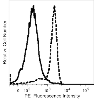
The L306.4 monoclonal antibody specifically recognizes CD58 which is also known as Lymphocyte function-associated antigen-3 (LFA-3) or Ag3. CD58 is a ~40 to 65 kDa cell-surface glycoprotein that belongs to the immunoglobulin superfamily. The CD58 antigen mediates cellular adhesion and participates in signal transduction when it binds to its ligand, the CD2 antigen. Cellular interactions regulated by the CD58/CD2 antigens are involved in the antigen-independent adhesion pathway and cytotoxic T lymphocyte (CTL) activity. The CD58 antigen has two isoforms. One isoform is anchored in the cell membrane by a glycophosphatidyl inositol tail, while the other is a type I transmembrane glycoprotein which has a transmembrane hydrophobic segment and a cytoplasmic segment composed of 12 amino acids. The CD58 antigen is widely distributed on cells of both hematopoietic and nonhematopoietic origin. The CD58 antigen is expressed on approximately 40% to 60% of peripheral blood lymphocytes, including CTL. It is also expressed on monocytes, granulocytes, B lymphoblastoid cell lines (such as JY and Daudi), platelets, vascular endothelium and smooth muscle, fibroblasts, and approximately 40% of bone marrow cells.
The antibody was conjugated to BD Horizon™ BV510 which is part of the BD Horizon Brilliant™ Violet family of dyes. With an Ex Max of 405-nm and Em Max at 510-nm, BD Horizon BV510 can be excited by the violet laser and detected in the BD Horizon V500 (525/50-nm) filter set. BD Horizon BV510 conjugates are useful for the detection of dim markers off the violet laser.

Development References (12)
-
Rincón J, Patarroyo M. Effect of antibodies from the T-cell (‘CD2 only’) and the NK/non-lineage (new panel only) sections on adhesion of Jurkat (T) cells to human erythrocytes. In: Knapp W. W. Knapp .. et al., ed. Leucocyte typing IV : white cell differentiation antigens. Oxford New York: Oxford University Press; 1989:718-720.
-
Carpén O, Dustin ML, Springer TA, Swafford JA, Beckett LA, Caulfield JP. Motility and ultrastructure of large granular lymphocytes on lipid bilayers reconstituted with adhesion receptors LFA-1, ICAM-1, and two isoforms of LFA-3.. J Cell Biol. 1991; 115(3):861-71. (Biology). View Reference
-
Dengler TJ, Hoffmann JC, Knolle P, et al. Structural and functional epitopes of the human adhesion receptor CD58 (LFA-3). Eur J Immunol. 1992; 22(11):2809-2817. (Biology). View Reference
-
Griffin H, Rowe M, Murray R, Brooks J, Rickinson A. Restoration of the LFA-3 adhesion pathway in Burkitt's lymphoma cells using an LFA-3 recombinant vaccinia virus: consequences for T cell recognition.. Eur J Immunol. 1992; 22(7):1741-8. (Biology). View Reference
-
Krensky AM, Robbins E, Springer TA, Burakoff SJ. LFA-1, LFA-2, and LFA-3 antigens are involved in CTL-target conjugation.. J Immunol. 1984; 132(5):2180-2. (Biology). View Reference
-
Krensky AM, Sanchez-Madrid F, Robbins E, Nagy JA, Springer TA, Burakoff SJ. The functional significance, distribution, and structure of LFA-1, LFA-2, and LFA-3: cell surface antigens associated with CTL-target interactions.. J Immunol. 1983; 131(2):611-6. (Biology). View Reference
-
Scheeren RA, Koopman G, Van der Baan S, Meijer CJ, Pals ST. Adhesion receptors involved in clustering of blood dendritic cells and T lymphocytes.. Eur J Immunol. 1991; 21(5):1101-5. (Biology). View Reference
-
Selvaraj P, Plunkett ML, Dustin M, Sanders ME, Shaw S, Springer TA. The T lymphocyte glycoprotein CD2 binds the cell surface ligand LFA-3.. Nature. 326(6111):400-3. (Biology). View Reference
-
Shaw S, Johnson JP. Cluster report: CD58. In: Knapp W. W. Knapp .. et al., ed. Leucocyte typing IV : white cell differentiation antigens. Oxford New York: Oxford University Press; 1989:714-716.
-
Shaw S, Luce GE, Quinones R, Gress RE, Springer TA, Sanders ME. Two antigen-independent adhesion pathways used by human cytotoxic T-cell clones.. Nature. 323(6085):262-4. (Biology). View Reference
-
Smith ME, Thomas JA. Cellular expression of lymphocyte function associated antigens and the intercellular adhesion molecule-1 in normal tissue.. J Clin Pathol. 1990; 43(11):893-900. (Clone-specific). View Reference
-
Springer TA. Adhesion receptors of the immune system. Nature. 1990; 346(6283):425-434. (Biology). View Reference
Please refer to Support Documents for Quality Certificates
Global - Refer to manufacturer's instructions for use and related User Manuals and Technical data sheets before using this products as described
Comparisons, where applicable, are made against older BD Technology, manual methods or are general performance claims. Comparisons are not made against non-BD technologies, unless otherwise noted.
For Research Use Only. Not for use in diagnostic or therapeutic procedures.