-
Reagents
- Flow Cytometry Reagents
-
Western Blotting and Molecular Reagents
- Immunoassay Reagents
-
Single-Cell Multiomics Reagents
- BD® OMICS-Guard Sample Preservation Buffer
- BD® AbSeq Assay
- BD® OMICS-One Immune Profiler Protein Panel
- BD® Single-Cell Multiplexing Kit
- BD Rhapsody™ ATAC-Seq Assays
- BD Rhapsody™ Whole Transcriptome Analysis (WTA) Amplification Kit
- BD Rhapsody™ TCR/BCR Next Multiomic Assays
- BD Rhapsody™ Targeted mRNA Kits
- BD Rhapsody™ Accessory Kits
-
Functional Assays
-
Microscopy and Imaging Reagents
-
Cell Preparation and Separation Reagents
-
- BD® OMICS-Guard Sample Preservation Buffer
- BD® AbSeq Assay
- BD® OMICS-One Immune Profiler Protein Panel
- BD® Single-Cell Multiplexing Kit
- BD Rhapsody™ ATAC-Seq Assays
- BD Rhapsody™ Whole Transcriptome Analysis (WTA) Amplification Kit
- BD Rhapsody™ TCR/BCR Next Multiomic Assays
- BD Rhapsody™ Targeted mRNA Kits
- BD Rhapsody™ Accessory Kits
- United States (English)
-
Change country/language
Old Browser
This page has been recently translated and is available in French now.
Looks like you're visiting us from {countryName}.
Would you like to stay on the current country site or be switched to your country?


Western blot analysis of OPA1 on a K-562 cell lysate (Human bone marrow myelogenous leukemia; ATCC CCL-243). Lane 1: 1:500, lane 2: 1000, lane 3: 1: 2000 dilution of the mouse anti- OPA1 antibody.

Immunofluorescence staining of COS-7 cells (African Green Monkey SV40 transformed kidney cells; ATCC CRL-1651).


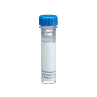
BD Transduction Laboratories™ Purified Mouse Anti-OPA1

BD Transduction Laboratories™ Purified Mouse Anti-OPA1

Regulatory Status Legend
Any use of products other than the permitted use without the express written authorization of Becton, Dickinson and Company is strictly prohibited.
Preparation And Storage
Recommended Assay Procedures
Western blot: Please refer to http://www.bdbiosciences.com/pharmingen/protocols/Western_Blotting.shtml
Product Notices
- Since applications vary, each investigator should titrate the reagent to obtain optimal results.
- Please refer to www.bdbiosciences.com/us/s/resources for technical protocols.
- Source of all serum proteins is from USDA inspected abattoirs located in the United States.
- Caution: Sodium azide yields highly toxic hydrazoic acid under acidic conditions. Dilute azide compounds in running water before discarding to avoid accumulation of potentially explosive deposits in plumbing.
Companion Products

.png?imwidth=320)
Three major GTP-binding protein families include trimeric and low molecular weight G-proteins, as well as a family of large proteins homologous to dynamin. The dynamin family contains proteins with diverse structure and function, but highly homologous N-terminal GTPase domains. A subgroup of the dynamin G-protein-binding family includes the mitochondrial proteins Drp1/Dnm1, Mgm1, and OPA1. The latter protein is mutated in dominant optic atrophy, a disease that involves loss of visual acuity and atrophy of the optic nerve. OPA1 is expressed in heart, brain, liver, and kidney. The sequence of OPA1 includes an N-terminal region that contains a mitochondrial targeting domain and three GTP-binding motifs. The overexpression of OPA1 in Cos-7 cells shows co-localization with cytochrome c in mitochondria, and leads to alterations in mitochondrial morphology from a characteristic tubuluar shape to a vesicular pattern. Thus, OPA1 may have roles in mitochondrial biogenesis that are critical for normal cell function.
This antibody is routinely tested by western blot analysis. Other applications were tested at BD Biosciences Pharmingen during antibody development only or reported in the literature.
Development References (3)
-
Alexander C, Votruba M, Pesch UE, et al. OPA1, encoding a dynamin-related GTPase, is mutated in autosomal dominant optic atrophy linked to chromosome 3q28. Nat Genet. 2000; 26(2):211-215. (Biology). View Reference
-
Delettre C, Lenaers G, Griffoin JM, et al. Nuclear gene OPA1, encoding a mitochondrial dynamin-related protein, is mutated in dominant optic atrophy. Nat Genet. 2000; 26(2):207-210. (Biology). View Reference
-
Misaka T, Miyashita T, Kubo Y. Primary structure of a dynamin-related mouse mitochondrial GTPase and its distribution in brain, subcellular localization, and effect on mitochondrial morphology. J Biol Chem. 2002; 277(18):15834-15842. (Biology). View Reference
Please refer to Support Documents for Quality Certificates
Global - Refer to manufacturer's instructions for use and related User Manuals and Technical data sheets before using this products as described
Comparisons, where applicable, are made against older BD Technology, manual methods or are general performance claims. Comparisons are not made against non-BD technologies, unless otherwise noted.
For Research Use Only. Not for use in diagnostic or therapeutic procedures.