-
Reagents
- Flow Cytometry Reagents
-
Western Blotting and Molecular Reagents
- Immunoassay Reagents
-
Single-Cell Multiomics Reagents
- BD® OMICS-Guard Sample Preservation Buffer
- BD® AbSeq Assay
- BD® OMICS-One Immune Profiler Protein Panel
- BD® Single-Cell Multiplexing Kit
- BD Rhapsody™ ATAC-Seq Assays
- BD Rhapsody™ Whole Transcriptome Analysis (WTA) Amplification Kit
- BD Rhapsody™ TCR/BCR Next Multiomic Assays
- BD Rhapsody™ Targeted mRNA Kits
- BD Rhapsody™ Accessory Kits
-
Functional Assays
-
Microscopy and Imaging Reagents
-
Cell Preparation and Separation Reagents
-
- BD® OMICS-Guard Sample Preservation Buffer
- BD® AbSeq Assay
- BD® OMICS-One Immune Profiler Protein Panel
- BD® Single-Cell Multiplexing Kit
- BD Rhapsody™ ATAC-Seq Assays
- BD Rhapsody™ Whole Transcriptome Analysis (WTA) Amplification Kit
- BD Rhapsody™ TCR/BCR Next Multiomic Assays
- BD Rhapsody™ Targeted mRNA Kits
- BD Rhapsody™ Accessory Kits
- United States (English)
-
Change country/language
Old Browser
This page has been recently translated and is available in French now.
Looks like you're visiting us from {countryName}.
Would you like to stay on the current country site or be switched to your country?


Western blot analysis of Mint3 on a RSV-3T3 cell lysate (left). Lane 1: 1:250, lane 2: 1:500, lane 3: 1:1000 dilution of the anti- Mint3 antibody. Immunofluorescent staining of SK-N-SH cells (right). Cells were seeded in a 384 well collagen coated Microplates (Material # 353962) at ~ 8,000 cells per well. After overnight incubation, cells were stained using the methanol fix/perm protocol (see Recommended Assay Procedure; Bioimaging protocol link) and the anti- Mint3-X11g antibody. The second step reagent was Alexa Fluor® 488 goat anti mouse Ig (Invitrogen)(pseudo colored green). Cell nuclei were counter stained with Hoechst 33342 (pseudo colored blue). The image was taken on a BD Pathway™ 855 or 435 Bioimager System using a 20x objective and merged using the BD AttoVison ™ software. This antibody also stained SK-N-SH, C6, U87 and U373 cells using both the Triton X100 and methanol fix/perm protocols (see Recommended Assay Procedure; Bioimaging protocol link).


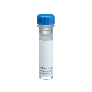
BD Transduction Laboratories™ Purified Mouse Anti-Mint3

Regulatory Status Legend
Any use of products other than the permitted use without the express written authorization of Becton, Dickinson and Company is strictly prohibited.
Preparation And Storage
Product Notices
- Since applications vary, each investigator should titrate the reagent to obtain optimal results.
- Caution: Sodium azide yields highly toxic hydrazoic acid under acidic conditions. Dilute azide compounds in running water before discarding to avoid accumulation of potentially explosive deposits in plumbing.
- Source of all serum proteins is from USDA inspected abattoirs located in the United States.
- Sodium azide is a reversible inhibitor of oxidative metabolism; therefore, antibody preparations containing this preservative agent must not be used in cell cultures nor injected into animals. Sodium azide may be removed by washing stained cells or plate-bound antibody or dialyzing soluble antibody in sodium azide-free buffer. Since endotoxin may also affect the results of functional studies, we recommend the NA/LE (No Azide/Low Endotoxin) antibody format, if available, for in vitro and in vivo use.
- This antibody has been developed and certified for the bioimaging application. However, a routine bioimaging test is not performed on every lot. Researchers are encouraged to titrate the reagent for optimal performance.
- Alexa Fluor® is a registered trademark of Molecular Probes, Inc., Eugene, OR.
- Triton is a trademark of the Dow Chemical Company.
- Please refer to www.bdbiosciences.com/us/s/resources for technical protocols.
Companion Products

.png?imwidth=320)
Munc18-1-interacting proteins, Mint1, Mint2, and Mint3, are members of the X11 family of proteins, which contain a phosphotyrosine-binding (PTB) domain and two PSD-95-DLG-ZO-1 (PDZ) domains. The PTB domain binds Asn-Pro-X-pTyr, a β-turn motif found on activated growth factor receptors and other signaling molecules. PDZ domains bind the C-terminus of proteins involved in receptor and channel clustering and protein localization in polarized cells. Both Mint1 and Mint2 are expressed primarily in the nervous system and may interact with Munc18-1 and syntaxin to form a multimeric complex that mediates appropriate docking/fusion of synaptic vesicles. In addition, both Mint1 and Mint2 may be involved in Alzheimer's disease, since Mint2 colocalizes with amyloid precursor protein (APP) and is found in neuritic plaques, while Mint1 can bind APP, and inhibits the processing of APP to the amyloid β peptide. Mint3 can also bind APP, but differs from Mint1 and Mint2 in its N-terminal region. It is also more widely expressed. Thus, Mint3 may function in signaling pathways, vesicle exocytosis, and/or protein targeting in a wide range of tissues. Mint3 has a calculated molecular weight of 61 kD, but reportedly is observed migrating at 86-89 kD.
Development References (3)
-
Borg JP, Straight SW, Kaech SM. Identification of an evolutionarily conserved heterotrimeric protein complex involved in protein targeting. J Biol Chem. 1998; 273(48):31633-31636. (Biology). View Reference
-
Borg JP, Yang Y, De Taddeo-Borg M, Margolis B, Turner RS. The X11alpha protein slows cellular amyloid precursor protein processing and reduces Abeta40 and Abeta42 secretion. J Biol Chem. 1998; 273(24):14761-14766. (Biology). View Reference
-
McLoughlin DM, Irving NG, Brownlees J, Brion JP, Leroy K, Miller CC. Mint2/X11-like colocalizes with the Alzheimer's disease amyloid precursor protein and is associated with neuritic plaques in Alzheimer's disease. Eur J Neurosci. 1999; 11(6):1988-1994. (Biology). View Reference
Please refer to Support Documents for Quality Certificates
Global - Refer to manufacturer's instructions for use and related User Manuals and Technical data sheets before using this products as described
Comparisons, where applicable, are made against older BD Technology, manual methods or are general performance claims. Comparisons are not made against non-BD technologies, unless otherwise noted.
For Research Use Only. Not for use in diagnostic or therapeutic procedures.