-
Reagents
- Flow Cytometry Reagents
-
Western Blotting and Molecular Reagents
- Immunoassay Reagents
-
Single-Cell Multiomics Reagents
- BD® OMICS-Guard Sample Preservation Buffer
- BD® AbSeq Assay
- BD® OMICS-One Immune Profiler Protein Panel
- BD® Single-Cell Multiplexing Kit
- BD Rhapsody™ ATAC-Seq Assays
- BD Rhapsody™ Whole Transcriptome Analysis (WTA) Amplification Kit
- BD Rhapsody™ TCR/BCR Next Multiomic Assays
- BD Rhapsody™ Targeted mRNA Kits
- BD Rhapsody™ Accessory Kits
-
Functional Assays
-
Microscopy and Imaging Reagents
-
Cell Preparation and Separation Reagents
-
- BD® OMICS-Guard Sample Preservation Buffer
- BD® AbSeq Assay
- BD® OMICS-One Immune Profiler Protein Panel
- BD® Single-Cell Multiplexing Kit
- BD Rhapsody™ ATAC-Seq Assays
- BD Rhapsody™ Whole Transcriptome Analysis (WTA) Amplification Kit
- BD Rhapsody™ TCR/BCR Next Multiomic Assays
- BD Rhapsody™ Targeted mRNA Kits
- BD Rhapsody™ Accessory Kits
- United States (English)
-
Change country/language
Old Browser
This page has been recently translated and is available in French now.
Looks like you're visiting us from {countryName}.
Would you like to stay on the current country site or be switched to your country?


Western blot analysis of NFAT-1 on a Jurkat cell lysate (Human T-cell leukemia; ATCC TIB-152). Lane 1: 1:2500, lane 2: 1:5000, lane 3: 1:10,000 dilution of the mouse anti- NFAT-1 antibody. NFAT-1 may be identified migrating between 97-135 kDa.

Immunofluorescence staining of Jurkat cells (Human T-cell leukemia; ATCC TIB-152).


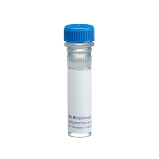
BD Transduction Laboratories™ Purified Mouse Anti- NFAT-1

BD Transduction Laboratories™ Purified Mouse Anti- NFAT-1

Regulatory Status Legend
Any use of products other than the permitted use without the express written authorization of Becton, Dickinson and Company is strictly prohibited.
Preparation And Storage
Recommended Assay Procedures
Western blot: Please refer to http://www.bdbiosciences.com/pharmingen/protocols/Western_Blotting.shtml
Product Notices
- Since applications vary, each investigator should titrate the reagent to obtain optimal results.
- Source of all serum proteins is from USDA inspected abattoirs located in the United States.
- Caution: Sodium azide yields highly toxic hydrazoic acid under acidic conditions. Dilute azide compounds in running water before discarding to avoid accumulation of potentially explosive deposits in plumbing.
- Please refer to www.bdbiosciences.com/us/s/resources for technical protocols.
Companion Products
.png?imwidth=320)


T cells are activated and induced to proliferate following binding of their respective antigen. The process of includes expression of genes that encode factors (i.e., cytokines) which regulate various cell types. Modulation of gene expression is conducted by an array of specific interactions between transcription factors and DNA. NFAT-1 (Nuclear Factor of Activated T cells) is a transcription factor that regulates expression of the interleukin-2 gene. Thus, NFAT-1 DNA binding activity is undetectable in resting cells, but increases during T-cell activation. NFAT-1, a protein of 921 amino acids, is part of an oligomeric transcription factor that also contains Fra-1 and JunB. NFAT-1 was initially described as a phosphoprotein and is dephosphorylated in activated T cells transformed with the leukemia virus HTLV-l.
Development References (5)
-
Boise LH, Petryniak B, Mao X, et al. The NFAT-1 DNA binding complex in activated T cells contains Fra-1 and JunB. Mol Cell Biol. 1993; 13(3):1911-1919. (Biology). View Reference
-
Good L, Maggirwar SB, Harhaj EW, Sun SC. Constitutive dephosphorylation and activation of a member of the nuclear factor of activated T cells, NF-AT1, in Tax-expressing and type I human T-cell leukemia virus-infected human T cells. J Biol Chem. 1997; 272(3):1425-1428. (Biology). View Reference
-
Grader-Beck T, van Puijenbroek AA, Nadler LM, Boussiotis VA. cAMP inhibits both Ras and Rap1 activation in primary human T lymphocytes, but only Ras inhibition correlates with blockade of cell cycle progression. Blood. 2003; 101(3):998-1006. (Biology: Western blot). View Reference
-
Kadereit S, Mohammad SF, Miller RE, et al. Reduced NFAT1 protein expression in human umbilical cord blood T lymphocytes. Blood. 1999; 94(9):3101-3107. (Biology: Western blot). View Reference
-
McCaffrey PG, Luo C, Kerppola TK, et al. Isolation of the cyclosporin-sensitive T cell transcription factor NFATp. Science. 1993; 262(5134):750-754. (Biology). View Reference
Please refer to Support Documents for Quality Certificates
Global - Refer to manufacturer's instructions for use and related User Manuals and Technical data sheets before using this products as described
Comparisons, where applicable, are made against older BD Technology, manual methods or are general performance claims. Comparisons are not made against non-BD technologies, unless otherwise noted.
For Research Use Only. Not for use in diagnostic or therapeutic procedures.