-
Reagents
- Flow Cytometry Reagents
-
Western Blotting and Molecular Reagents
- Immunoassay Reagents
-
Single-Cell Multiomics Reagents
- BD® OMICS-Guard Sample Preservation Buffer
- BD® AbSeq Assay
- BD® OMICS-One Immune Profiler Protein Panel
- BD® Single-Cell Multiplexing Kit
- BD Rhapsody™ ATAC-Seq Assays
- BD Rhapsody™ Whole Transcriptome Analysis (WTA) Amplification Kit
- BD Rhapsody™ TCR/BCR Next Multiomic Assays
- BD Rhapsody™ Targeted mRNA Kits
- BD Rhapsody™ Accessory Kits
-
Functional Assays
-
Microscopy and Imaging Reagents
-
Cell Preparation and Separation Reagents
-
- BD® OMICS-Guard Sample Preservation Buffer
- BD® AbSeq Assay
- BD® OMICS-One Immune Profiler Protein Panel
- BD® Single-Cell Multiplexing Kit
- BD Rhapsody™ ATAC-Seq Assays
- BD Rhapsody™ Whole Transcriptome Analysis (WTA) Amplification Kit
- BD Rhapsody™ TCR/BCR Next Multiomic Assays
- BD Rhapsody™ Targeted mRNA Kits
- BD Rhapsody™ Accessory Kits
- United States (English)
-
Change country/language
Old Browser
This page has been recently translated and is available in French now.
Looks like you're visiting us from {countryName}.
Would you like to stay on the current country site or be switched to your country?
![Purified Mouse Anti- PKA [RI]](/content/dam/bdb/products/global/reagents/microscopy-imaging-reagents/immunofluorescence-reagents/610xxx/6101xx/610166_base/P19920_610166_image1.png)
![Purified Mouse Anti- PKA [RI]](/content/dam/bdb/products/global/reagents/microscopy-imaging-reagents/immunofluorescence-reagents/610xxx/6101xx/610166_base/P19920_610165_2.png)

![Purified Mouse Anti- PKA [RI]](/content/dam/bdb/products/global/reagents/microscopy-imaging-reagents/immunofluorescence-reagents/610xxx/6101xx/610166_base/P19920_610166_image1.png)
Western blot analysis of PKA [RI] on a human endothelial lysate. Lane 1: 1:250, lane 2: 1:500, lane 3: 1:1000 dilution of the PKA [RI] antibody.
![Purified Mouse Anti- PKA [RI]](/content/dam/bdb/products/global/reagents/microscopy-imaging-reagents/immunofluorescence-reagents/610xxx/6101xx/610166_base/P19920_610165_2.png)
Immunofluorescent staining of A549 (ATCC CCL-185) cells. Cells were seeded in a 96 well imaging plate (Cat. No. 353219) at ~ 10 000 cells per well. After overnight incubation, cells were stained using the alcohol perm protocol and the anti-PKA [RI] antibody. The second step reagent was FITC goat anti mouse Ig (Cat. No. 554001). The image was taken on a BD Pathway™ 855 Bioimager using a 20x objective. This antibody also stained HeLa (ATCC CCL-2) and U-2 OS (ATCC HTB-96) cells using both the Triton™ X-100 and alcohol perm protocol (see Recommended Assay Procedure).

![Purified Mouse Anti- PKA [RI]](/content/dam/bdb/products/global/reagents/microscopy-imaging-reagents/immunofluorescence-reagents/610xxx/6101xx/610166_base/P19920_610166_image1.png)
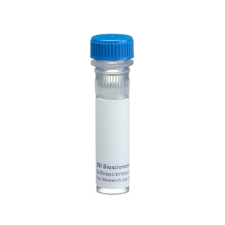
BD Transduction Laboratories™ Purified Mouse Anti- PKA [RI]
![Purified Mouse Anti- PKA [RI]](/content/dam/bdb/products/global/reagents/microscopy-imaging-reagents/immunofluorescence-reagents/610xxx/6101xx/610166_base/P19920_610165_2.png)
BD Transduction Laboratories™ Purified Mouse Anti- PKA [RI]

Regulatory Status Legend
Any use of products other than the permitted use without the express written authorization of Becton, Dickinson and Company is strictly prohibited.
Preparation And Storage
Recommended Assay Procedures
Bioimaging
1. Seed the cells in appropriate culture medium at ~10,000 cells per well in a BD Falcon™ 96-well Imaging Plate (Cat. No. 353219) and culture overnight.
2. Remove the culture medium from the wells, and fix the cells by adding 100 μl of BD Cytofix™ Fixation Buffer (Cat. No. 554655) to each well. Incubate for 10 minutes at room temperature (RT).
3. Remove the fixative from the wells, and permeabilize the cells using either BD Perm Buffer III, 90% methanol, or Triton™ X-100:
a. Add 100 μl of -20°C 90% methanol or Perm Buffer III (Cat. No. 558050) to each well and incubate for 5 minutes at RT.
OR
b. Add 100 μl of 0.1% Triton™ X-100 to each well and incubate for 5 minutes at RT.
4. Remove the permeabilization buffer, and wash the wells twice with 100 μl of 1× PBS.
5. Remove the PBS, and block the cells by adding 100 μl of BD Pharmingen™ Stain Buffer (FBS) (Cat. No. 554656) to each well. Incubate for 30 minutes at RT.
6. Remove the blocking buffer and add 50 μl of the optimally titrated primary antibody (diluted in Stain Buffer) to each well, and incubate for 1 hour at RT.
7. Remove the primary antibody, and wash the wells three times with 100 μl of 1× PBS.
8. Remove the PBS, and add the second step reagent at its optimally titrated concentration in 50 μl to each well, and incubate in the dark for 1 hour at RT.
9. Remove the second step reagent, and wash the wells three times with 100 μl of 1× PBS.
10. Remove the PBS, and counter-stain the nuclei by adding 200 μl per well of 2 μg/ml Hoechst 33342 (e.g., Sigma-Aldrich Cat. No. B2261) in 1× PBS to each well at least 15 minutes before imaging.
11. View and analyze the cells on an appropriate imaging instrument.
Bioimaging: For more detailed information please refer to http://www.bdbiosciences.com/support/resources/protocols/ceritifed_reagents.jsp
Western blot: For more detailed information please refer to http://www.bdbiosciences.com/pharmingen/protocols/Western_Blotting.shtml
Product Notices
- Since applications vary, each investigator should titrate the reagent to obtain optimal results.
- Please refer to www.bdbiosciences.com/us/s/resources for technical protocols.
- This antibody has been developed and certified for the bioimaging application. However, a routine bioimaging test is not performed on every lot. Researchers are encouraged to titrate the reagent for optimal performance.
- Caution: Sodium azide yields highly toxic hydrazoic acid under acidic conditions. Dilute azide compounds in running water before discarding to avoid accumulation of potentially explosive deposits in plumbing.
- Source of all serum proteins is from USDA inspected abattoirs located in the United States.
- Triton is a trademark of the Dow Chemical Company.
Companion Products


.png?imwidth=320)


cAMP-dependent Protein Kinase (PKA) is composed of two distinct subunits: catalytic (C) and regulatory (R). Four regulatory subunits have been identified: RIα, RIß, RIIα, and RIIß. These subunits define type I and II cAMP-dependent protein kinases. Following binding of cAMP, the regulatory subunits dissociate from the catalytic subunits, rendering the enzyme active. Type I and type II holoenzymes have three potential C subunits (Cα, Cß, or Cγ). Type II PKA can be distinguished by autophosphorylation of the R-subunits, while type I PKA binds Mg/ATP with high affinity. Most cells express both type I and type II PKAs. Although the Rα isoforms are ubiquitously expressed, the Rß isoforms are predominant in nervous and adipose tissues. The levels of expression of the different subunits vary according to cell and tissue type.
Development References (5)
-
Chen W, Yu YL, Lee SF, et al. CREB is one component of the binding complex of the Ces-2/E2A-HLF binding element and is an integral part of the interleukin-3 survival signal. Mol Cell Biol. 2001; 21(14):4636-4646. (Clone-specific: Flow cytometry). View Reference
-
Cho-Chung YS. Role of cyclic AMP receptor proteins in growth, differentiation, and suppression of malignancy: new approaches to therapy. Cancer Res. 1990; 50(22):7093-7100. (Biology). View Reference
-
Dohrman DP, Diamond I, Gordon AS. Ethanol causes translocation of cAMP-dependent protein kinase catalytic subunit to the nucleus. Proc Natl Acad Sci U S A. 1996; 93(19):10217-10221. (Clone-specific: Immunofluorescence). View Reference
-
Orellana SA, Marfella-Scivittaro C. Distinctive cyclic AMP-dependent protein kinase subunit localization is associated with cyst formation and loss of tubulogenic capacity in Madin-Darby canine kidney cell clones. J Biol Chem. 2000; 275(28):21233-21240. (Clone-specific: Immunofluorescence, Western blot). View Reference
-
Taylor SS, Buechler JA, Yonemoto W. cAMP-dependent protein kinase: framework for a diverse family of regulatory enzymes. Annu Rev Biochem. 1990; 59:971-1005. (Biology). View Reference
Please refer to Support Documents for Quality Certificates
Global - Refer to manufacturer's instructions for use and related User Manuals and Technical data sheets before using this products as described
Comparisons, where applicable, are made against older BD Technology, manual methods or are general performance claims. Comparisons are not made against non-BD technologies, unless otherwise noted.
For Research Use Only. Not for use in diagnostic or therapeutic procedures.