-
Reagents
- Flow Cytometry Reagents
-
Western Blotting and Molecular Reagents
- Immunoassay Reagents
-
Single-Cell Multiomics Reagents
- BD® OMICS-Guard Sample Preservation Buffer
- BD® AbSeq Assay
- BD® OMICS-One Immune Profiler Protein Panel
- BD® Single-Cell Multiplexing Kit
- BD Rhapsody™ ATAC-Seq Assays
- BD Rhapsody™ Whole Transcriptome Analysis (WTA) Amplification Kit
- BD Rhapsody™ TCR/BCR Next Multiomic Assays
- BD Rhapsody™ Targeted mRNA Kits
- BD Rhapsody™ Accessory Kits
-
Functional Assays
-
Microscopy and Imaging Reagents
-
Cell Preparation and Separation Reagents
-
- BD® OMICS-Guard Sample Preservation Buffer
- BD® AbSeq Assay
- BD® OMICS-One Immune Profiler Protein Panel
- BD® Single-Cell Multiplexing Kit
- BD Rhapsody™ ATAC-Seq Assays
- BD Rhapsody™ Whole Transcriptome Analysis (WTA) Amplification Kit
- BD Rhapsody™ TCR/BCR Next Multiomic Assays
- BD Rhapsody™ Targeted mRNA Kits
- BD Rhapsody™ Accessory Kits
- United States (English)
-
Change country/language
Old Browser
This page has been recently translated and is available in French now.
Looks like you're visiting us from {countryName}.
Would you like to stay on the current country site or be switched to your country?


Multicolor flow cytometric analysis of Foxp3 expression in mouse splenocytes. C57BL/6 mouse spleen cells were treated with BD Pharm Lyse™ Lysing Buffer (Cat. No. 555899) to lyse erythrocytes. The leucocytes were washed, fixed and permeabilized using the Transcription Factor Buffer Set (Cat. No. 562574/562725). The cells were then stained with FITC Rat Anti-Mouse CD4 antibody (Cat. No. 553729), BD Horizon™ BUV395 Rat Anti-Mouse CD25 antibody (Cat. No. 564022) and with either BD Horizon™ BV421 Mouse IgG1, κ Isotype Control (Cat. No. 562438; Top Plots) or BD Horizon™ BV421 Mouse Anti-Foxp3 antibody (Cat. No. 567458; Bottom plots) at 0.5 µg/test. Left Plots: Bivariate pseudocolor density plots showing the correlated expression of CD4 versus Foxp3 (or Ig Isotype control staining) were derived from gated events with the forward and side light-scatter characteristics of intact cells. Right Plots: Bivariate pseudocolor density plots showing the correlated expression of Foxp3 (or Ig Isotype control staining) versus CD25 were derived from CD4 positive-gated events with the light-scatter characteristics of intact cells. Flow cytometric analysis was performed using a BD LSRFortessa™ X-20 Flow Cytometer System and FlowJo™ software. Data shown on this Technical Data Sheet are not lot specific.


BD Horizon™ BV421 Mouse Anti-Mouse FoxP3

Regulatory Status Legend
Any use of products other than the permitted use without the express written authorization of Becton, Dickinson and Company is strictly prohibited.
Preparation And Storage
Recommended Assay Procedures
BD® CompBeads can be used as surrogates to assess fluorescence spillover (compensation). When fluorochrome conjugated antibodies are bound to BD® CompBeads, they have spectral properties very similar to cells. However, for some fluorochromes there can be small differences in spectral emissions compared to cells, resulting in spillover values that differ when compared to biological controls. It is strongly recommended that when using a reagent for the first time, users compare the spillover on cells and BD® CompBeads to ensure that BD® CompBeads are appropriate for your specific cellular application.
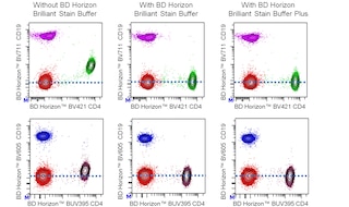
For optimal and reproducible results, BD Horizon Brilliant Stain Buffer should be used anytime BD Horizon Brilliant dyes are used in a multicolor flow cytometry panel. Fluorescent dye interactions may cause staining artifacts which may affect data interpretation. The BD Horizon Brilliant Stain Buffer was designed to minimize these interactions. When BD Horizon Brilliant Stain Buffer is used in in the multicolor panel, it should also be used in the corresponding compensation controls for all dyes to achieve the most accurate compensation. For the most accurate compensation, compensation controls created with either cells or beads should be exposed to BD Horizon Brilliant Stain Buffer for the same length of time as the corresponding multicolor panel. More information can be found in the Technical Data Sheet of the BD Horizon Brilliant Stain Buffer (Cat. No. 563794/566349) or the BD Horizon Brilliant Stain Buffer Plus (Cat. No. 566385).
Product Notices
- Please refer to www.bdbiosciences.com/us/s/resources for technical protocols.
- Source of all serum proteins is from USDA inspected abattoirs located in the United States.
- Caution: Sodium azide yields highly toxic hydrazoic acid under acidic conditions. Dilute azide compounds in running water before discarding to avoid accumulation of potentially explosive deposits in plumbing.
- Since applications vary, each investigator should titrate the reagent to obtain optimal results.
- For fluorochrome spectra and suitable instrument settings, please refer to our Multicolor Flow Cytometry web page at www.bdbiosciences.com/colors.
- An isotype control should be used at the same concentration as the antibody of interest.
- BD Horizon Brilliant Violet 421 is covered by one or more of the following US patents: 8,158,444; 8,362,193; 8,575,303; 8,354,239.
- BD Horizon Brilliant Stain Buffer is covered by one or more of the following US patents: 8,110,673; 8,158,444; 8,575,303; 8,354,239.
- Please refer to http://regdocs.bd.com to access safety data sheets (SDS).
- Pacific Blue™ is a trademark of Life Technologies Corporation.
Companion Products


The 3G3 monoclonal antibody specifically binds to Forkhead box protein P3 (Foxp3) which is also known as Scurfin and JM2. Foxp3 is a 50-55 kDa protein that is encoded by Foxp3 (forkhead box P3) which belongs to the forkhead/winged helix family of transcriptional regulators. FoxP3 contains a single C2H2-type zinc-finger motif, a leucine-zipper region, and a C-terminal forkhead DNA-binding domain. Foxp3 is expressed by natural (nTreg) and induced/adaptive (iTreg) T regulatory cells. Foxp3 is a key transcription factor for Treg cell development and regulatory function. Treg cells play crucial roles in maintaining immune homeostasis by enforcing immunological tolerance to self-antigens and by suppressing excessive responses made by other immune cells to foreign antigens. Ectopic expression of Foxp3 in conventional T cells is sufficient to induce suppressive activity, repress the production of cytokines such as IL2 and IFN-γ, and upregulate Treg cell-associated molecules such as CTLA4/CD152 and GITR/TNFRSF18. A mutation in Foxp3 causes a lack of functional Tregs in Scurfy (sf) mice which develop a systemic disease state with autoimmune manifestations. The 3G3 antibody reportedly binds to the N-terminus of Foxp3.

Development References (5)
-
Brunkow ME, Jeffery EW, Hjerrild KA, et al. Disruption of a new forkhead/winged-helix protein, scurfin, results in the fatal lymphoproliferative disorder of the scurfy mouse. Nat Genet. 2001; 27(1):68-73. (Biology). View Reference
-
Gavin MA, Torgerson TR, Houston E, et al. Single-cell analysis of normal and FOXP3-mutant human T cells: FOXP3 expression without regulatory T cell development. Proc Natl Acad Sci U S A. 2006; 103(17):6659-6664. (Immunogen: Flow cytometry). View Reference
-
Hadaschik EN, Wei X, Leiss H, et al. Regulatory T cell-deficient scurfy mice develop systemic autoimmune features resembling lupus-like disease. Arthritis Res Ther. 2015; 17(35):1-12. (Biology). View Reference
-
Law JP, Hirschkorn DF, Owen RE, Biswas HH, Norris PJ, Lanteri MC. The importance of Foxp3 antibody and fixation/permeabilization buffer combinations in identifying CD4+CD25+Foxp3+ regulatory T cells. Cytometry A. 2009; 75(12):1040-1050. (Clone-specific: Flow cytometry). View Reference
-
Mailer RKW. Alternative Splicing of FOXP3-Virtue and Vice. Front Immunol. 2018; 9(530):1-11. (Clone-specific). View Reference
Please refer to Support Documents for Quality Certificates
Global - Refer to manufacturer's instructions for use and related User Manuals and Technical data sheets before using this products as described
Comparisons, where applicable, are made against older BD Technology, manual methods or are general performance claims. Comparisons are not made against non-BD technologies, unless otherwise noted.
For Research Use Only. Not for use in diagnostic or therapeutic procedures.