-
Reagents
- Flow Cytometry Reagents
-
Western Blotting and Molecular Reagents
- Immunoassay Reagents
-
Single-Cell Multiomics Reagents
- BD® OMICS-Guard Sample Preservation Buffer
- BD® AbSeq Assay
- BD® OMICS-One Immune Profiler Protein Panel
- BD® Single-Cell Multiplexing Kit
- BD Rhapsody™ ATAC-Seq Assays
- BD Rhapsody™ Whole Transcriptome Analysis (WTA) Amplification Kit
- BD Rhapsody™ TCR/BCR Next Multiomic Assays
- BD Rhapsody™ Targeted mRNA Kits
- BD Rhapsody™ Accessory Kits
-
Functional Assays
-
Microscopy and Imaging Reagents
-
Cell Preparation and Separation Reagents
-
- BD® OMICS-Guard Sample Preservation Buffer
- BD® AbSeq Assay
- BD® OMICS-One Immune Profiler Protein Panel
- BD® Single-Cell Multiplexing Kit
- BD Rhapsody™ ATAC-Seq Assays
- BD Rhapsody™ Whole Transcriptome Analysis (WTA) Amplification Kit
- BD Rhapsody™ TCR/BCR Next Multiomic Assays
- BD Rhapsody™ Targeted mRNA Kits
- BD Rhapsody™ Accessory Kits
- United States (English)
-
Change country/language
Old Browser
This page has been recently translated and is available in French now.
Looks like you're visiting us from {countryName}.
Would you like to stay on the current country site or be switched to your country?


Two-color image analysis of CD3 Molecular Complex expression by cells within C57BL/6 mouse spleen. Mouse spleen cryosections (5 µm) were fixed with BD Cytofix™ Fixation Buffer (Cat. No. 554655), blocked with 5% goat serum and 1% BSA diluted in 1× PBS, and stained with BD Horizon™ BV480 Rat Anti-Mouse CD3 Molecular Complex antibody (Cat. No. 565642; pseudo-colored green) and BD Horizon™ BV421 Rat Anti-Mouse CD45R/B220 antibody (Cat. No. 562922; pseudo-colored red). Slides were mounted with ProLong® Gold and the images were captured on a standard epifluorescence microscope. Original magnification, 20×.


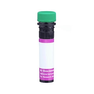
BD Horizon™ BV480 Rat Anti-Mouse CD3 Molecular Complex

Regulatory Status Legend
Any use of products other than the permitted use without the express written authorization of Becton, Dickinson and Company is strictly prohibited.
Preparation And Storage
Recommended Assay Procedures
For optimal and reproducible results, BD Horizon Brilliant Stain Buffer should be used anytime two or more BD Horizon Brilliant dyes are used in the same experiment. Fluorescent dye interactions may cause staining artifacts which may affect data interpretation. The BD Horizon Brilliant Stain Buffer was designed to minimize these interactions. More information can be found in the Technical Data Sheet of the BD Horizon Brilliant Stain Buffer (Cat. No. 563794/566349).
For Immunofluorescence Applications:
The use of a mounting reagent (eg, ProLong® Gold) is highly recommended to maximize the photostability of BV480. For confocal microscopy systems, a 440 nm laser is the optimal excitation source and the recommended emission filter is a 485/20 nm bandpass filter.
For epifluorescence microscopes with broad spectrum excitation sources, the recommended excitation and emission filters are 445/20 nm and 485/20 nm bandpass filters, respectively. For specific multicolor imaging applications, the exact filter configurations should be optimized by the end user. For additional instrument/filter configuration information, please visit http://www.bdbiosciences.com/research/cellularimaging.
Product Notices
- Since applications vary, each investigator should titrate the reagent to obtain optimal results.
- An isotype control should be used at the same concentration as the antibody of interest.
- Source of all serum proteins is from USDA inspected abattoirs located in the United States.
- Caution: Sodium azide yields highly toxic hydrazoic acid under acidic conditions. Dilute azide compounds in running water before discarding to avoid accumulation of potentially explosive deposits in plumbing.
- This antibody has been developed for the immunofluorescence imaging application. However, the antibody is routinely QC tested by flow cytometric analysis. Researchers are encouraged to titrate the reagent for optimal performance.
- ProLong® is a registered trademark of Thermo Fisher Scientific, Inc. Waltham, MA.
- For fluorochrome spectra and suitable instrument settings, please refer to our Multicolor Flow Cytometry web page at www.bdbiosciences.com/colors.
- BD Horizon Brilliant Violet 480 is covered by one or more of the following US patents: 8,575,303; 8,354,239.
- BD Horizon Brilliant Stain Buffer is covered by one or more of the following US patents: 8,110,673; 8,158,444; 8,575,303; 8,354,239.
- Please refer to www.bdbiosciences.com/us/s/resources for technical protocols.
Companion Products


The 17A2 monoclonal antibody specifically binds to the T-cell receptor-associated CD3 complex that is expressed on many thymocytes and mature T lymphocytes. Plate-bound 17A2 antibody has been reported to induce IL-2 production by cultured T cells in the absence of accessory cells. The binding of the 17A2 antibody to T cells can be blocked by the anti-CD3e mAb 145-2C11 (Cat. No. 557306/553058/550275). This suggests that the 17A2 antibody recognizes an epitope of the CD3 epsilon chain. In vivo treatment with 17A2 antibody has been reported to partially deplete T lymphocytes and temporarily down-modulates CD3 expression on T cells.
The antibody was conjugated to BD Horizon BV480 which is part of the BD Horizon Brilliant™ Violet family of dyes. With an Ex Max of 436-nm and Em Max at 478-nm, BD Horizon BV480 can be excited by the violet laser and detected in the BD Horizon BV510 (525/40-nm) filter set. BV480 has less spillover into the BV605 detector and, in general, is brighter than BV510.

Development References (3)
-
Miescher GC, Schreyer M, MacDonald HR. Production and characterization of a rat monoclonal antibody against the murine CD3 molecular complex. Immunol Lett. 1989; 23(2):113-118. (Immunogen: Cytotoxicity, Flow cytometry, Functional assay, Immunohistochemistry, Immunoprecipitation, Stimulation). View Reference
-
Mysliwietz J, Thierfelder S. Antilymphocytic antibodies and marrow transplantation. XII. Suppression of graft-versus-host disease by T-cell-modulating and depleting antimouse CD3 antibody is most effective when preinjected in the marrow recipient. Blood. 1992; 80(10):2661-2667. (Clone-specific: Depletion, Flow cytometry, Functional assay, In vivo exacerbation). View Reference
-
Wu L, Antica M, Johnson GR, Scollay R, Shortman K. Developmental potential of the earliest precursor cells from the adult mouse thymus. J Exp Med. 1991; 174(6):1617-1627. (Clone-specific: Cell separation, Depletion). View Reference
Please refer to Support Documents for Quality Certificates
Global - Refer to manufacturer's instructions for use and related User Manuals and Technical data sheets before using this products as described
Comparisons, where applicable, are made against older BD Technology, manual methods or are general performance claims. Comparisons are not made against non-BD technologies, unless otherwise noted.
For Research Use Only. Not for use in diagnostic or therapeutic procedures.