-
Reagents
- Flow Cytometry Reagents
-
Western Blotting and Molecular Reagents
- Immunoassay Reagents
-
Single-Cell Multiomics Reagents
- BD® OMICS-Guard Sample Preservation Buffer
- BD® AbSeq Assay
- BD® OMICS-One Immune Profiler Protein Panel
- BD® Single-Cell Multiplexing Kit
- BD Rhapsody™ ATAC-Seq Assays
- BD Rhapsody™ Whole Transcriptome Analysis (WTA) Amplification Kit
- BD Rhapsody™ TCR/BCR Next Multiomic Assays
- BD Rhapsody™ Targeted mRNA Kits
- BD Rhapsody™ Accessory Kits
-
Functional Assays
-
Microscopy and Imaging Reagents
-
Cell Preparation and Separation Reagents
-
- BD® OMICS-Guard Sample Preservation Buffer
- BD® AbSeq Assay
- BD® OMICS-One Immune Profiler Protein Panel
- BD® Single-Cell Multiplexing Kit
- BD Rhapsody™ ATAC-Seq Assays
- BD Rhapsody™ Whole Transcriptome Analysis (WTA) Amplification Kit
- BD Rhapsody™ TCR/BCR Next Multiomic Assays
- BD Rhapsody™ Targeted mRNA Kits
- BD Rhapsody™ Accessory Kits
- United States (English)
-
Change country/language
Old Browser
This page has been recently translated and is available in French now.
Looks like you're visiting us from {countryName}.
Would you like to stay on the current country site or be switched to your country?


Two-color image analysis of CD45R/B220 expression by cells within C57BL/6 mouse spleen. Mouse spleen cryosections (5 µm) were fixed with BD Cytofix™ Fixation Buffer (Cat. No. 554655), blocked with 5% goat serum and 1% BSA diluted in 1× PBS, and stained with BD Horizon™ BV480 Rat Anti-Mouse CD45R/B220 antibody (Cat. No. 565631, pseudo-colored green) and Alexa Fluor® 647 Rat Anti-Mouse CD4 antibody (Cat. No. 557681, pseudo-colored red). Slides were mounted with ProLong® Gold and the images were captured on a standard epifluorescence microscope. Acquired at 20× magnification.


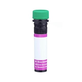
BD Horizon™ BV480 Rat Anti-Mouse CD45R/B220

Regulatory Status Legend
Any use of products other than the permitted use without the express written authorization of Becton, Dickinson and Company is strictly prohibited.
Preparation And Storage
Recommended Assay Procedures
For optimal and reproducible results, BD Horizon Brilliant Stain Buffer should be used anytime two or more BD Horizon Brilliant dyes are used in the same experiment. Fluorescent dye interactions may cause staining artifacts which may affect data interpretation. The BD Horizon Brilliant Stain Buffer was designed to minimize these interactions. More information can be found in the Technical Data Sheet of the BD Horizon Brilliant Stain Buffer (Cat. No. 563794/566349).
For Immunofluorescence Applications:
The use of a mounting reagent (eg, ProLong® Gold) is highly recommended to maximize the photostability of BV480. For confocal microscopy systems, a 440 nm laser is the optimal excitation source and the recommended emission filter is a 485/20 nm bandpass filter.
For epifluorescence microscopes with broad spectrum excitation sources, the recommended excitation and emission filters are 445/20 nm and 485/20 nm bandpass filters, respectively. For specific multicolor imaging applications, the exact filter configurations should be optimized by the end user. For additional instrument/filter configuration information, please visit http://www.bdbiosciences.com/research/cellularimaging.
Product Notices
- Since applications vary, each investigator should titrate the reagent to obtain optimal results.
- An isotype control should be used at the same concentration as the antibody of interest.
- Source of all serum proteins is from USDA inspected abattoirs located in the United States.
- Caution: Sodium azide yields highly toxic hydrazoic acid under acidic conditions. Dilute azide compounds in running water before discarding to avoid accumulation of potentially explosive deposits in plumbing.
- This antibody has been developed for the immunofluorescence imaging application. However, the antibody is routinely QC tested by flow cytometric analysis. Researchers are encouraged to titrate the reagent for optimal performance.
- BD Horizon Brilliant Violet 480 is covered by one or more of the following US patents: 8,575,303; 8,354,239.
- ProLong® is a registered trademark of Thermo Fisher Scientific, Inc. Waltham, MA.
- For fluorochrome spectra and suitable instrument settings, please refer to our Multicolor Flow Cytometry web page at www.bdbiosciences.com/colors.
- BD Horizon Brilliant Stain Buffer is covered by one or more of the following US patents: 8,110,673; 8,158,444; 8,575,303; 8,354,239.
- Alexa Fluor® is a registered trademark of Molecular Probes, Inc., Eugene, OR.
- Please refer to www.bdbiosciences.com/us/s/resources for technical protocols.
Companion Products


The RA3-6B2 monoclonal antibody specifically binds to an epitope on the extracellular domain of the transmembrane CD45 glycoprotein which is dependent upon the expression of exon A and specific carbohydrate residues. It is expressed on B lymphocytes at all stages from pro-B through mature and activated B cell, but it is decreased on plasma cells and a subset of memory B cells. The levels of CD45R expression on the B-cell lineage appear to be developmentally regulated. It is also reportedly found on the abnormal T cells involved in the lymphadenopathy of lpr/lpr and gld/gld mutant mice, on lytically active subsets of lymphokine-activated killer cells (NK cells and non-MHC-restricted CTL), on apoptotic T lymphocytes of mice injected with bacterial superantigen, on a population of NK-cell precursors in the bone marrow, and on B-lymphocyte, T-lymphocyte, and macrophage progenitors in fetal liver. The CD45R antigen has been reported not to be on hematopoietic stem cells, naive T lymphocytes, or MHC-restricted CTL. CD45 is a member of the Protein Tyrosine Phosphatase (PTP) family: Its intracellular (COOH-terminal) region contains two PTP catalytic domains, and the extracellular region is highly variable due to alternative splicing of exons 4, 5, and 6 (designated A, B, and C, respectively), plus differing levels of glycosylation. The CD45 isoforms detected in the mouse are cell type-, maturation, and activation state-specific. The CD45 isoforms play complex roles in T-cell and B-cell antigen receptor signal transduction. CD45R is commonly used as a pan B-cell marker; however, CD19 expression, detectable by the rat anti-mouse CD19 antibody (clone 1D3), is reported to be more restricted to the B-cell lineage. The rat anti-mouse CD45R antibody (clone RA3-6B2) has been reported to enhance isotype switching during in vitro B-cell responses and to inhibit in vivo B-cell responses. Cross-reaction of the RA3-6B2 clone with activated human T lymphocytes has also been reportedly observed.
The antibody was conjugated to BD Horizon BV480 which is part of the BD Horizon Brilliant™ Violet family of dyes. With an Ex Max of 436-nm and Em Max at 478-nm, BD Horizon BV480 can be excited by the violet laser and detected in the BD Horizon BV510 (525/40-nm) filter set. BV480 has less spillover into the BV605 detector and, in general, is brighter than BV510.

Development References (17)
-
Allman DM, Ferguson SE, Cancro MP. Peripheral B cell maturation. I. Immature peripheral B cells in adults are heat-stable antigenhi and exhibit unique signaling characteristics. J Immunol. 1992; 149(8):2533-2540. (Clone-specific: Flow cytometry). View Reference
-
Asensi V, Kimeno K, Kawamura I, Sakumoto M, Nomoto K. Treatment of autoimmune MRL/lpr mice with anti-B220 monoclonal antibody reduces the level of anti-DNA antibodies and lymphadenopathies. Immunology. 1989; 68(2):204-208. (Clone-specific: Flow cytometry, In vivo exacerbation). View Reference
-
Ballas ZK, Rasmussen W. Lymphokine-activated killer cells. VII. IL-4 induces an NK1.1+CD8 alpha+beta- TCR-alpha beta B220+ lymphokine-activated killer subset. J Immunol. 1993; 150(1):17-30. (Clone-specific: Flow cytometry, Fluorescence activated cell sorting). View Reference
-
Bleesing JJ, Morrow MR, Uzel G, Fleisher TA. Human T cell activation induces the expression of a novel CD45 isoform that is analogous to murine B220 and is associated with altered O-glycan synthesis and onset of apoptosis. Cell Immunol. 2001; 213(1):72-81. (Clone-specific: Flow cytometry). View Reference
-
Coffman RL. Surface antigen expression and immunoglobulin gene rearrangement during mouse pre-B cell development. Immunol Rev. 1982; 69:5-23. (Immunogen: Blocking, Flow cytometry, Immunofluorescence, Immunoprecipitation). View Reference
-
Domiati-Saad R, Ogle EW, Justement LB. Administration of anti-CD45 mAb specific for a B cell-restricted epitope abrogates the B cell response to a T-dependent antigen in vivo. J Immunol. 1993; 151(11):5936-5947. (Clone-specific: In vivo exacerbation). View Reference
-
Driver DJ, McHeyzer-Williams LJ, Cool M, Stetson DB, McHeyzer-Williams MG. Development and maintenance of a B220- memory B cell compartment. J Immunol. 2001; 167(3):1393-1405. (Clone-specific: Flow cytometry, Fluorescence activated cell sorting, Fluorescence microscopy, Immunofluorescence). View Reference
-
George A, Rath S, Shroff KE, Wang M, Durdik JM. Ligation of CD45 on B cells can facilitate production of secondary Ig isotypes. J Immunol. 1994; 152(3):1014-1021. (Clone-specific: Functional assay). View Reference
-
Hardy RR, Carmack CE, Shinton SA, Kemp JD, Hayakawa K. Resolution and characterization of pro-B and pre-pro-B cell stages in normal mouse bone marrow. J Exp Med. 1991; 173(5):1213-1225. (Clone-specific: Flow cytometry, Fluorescence activated cell sorting, Immunofluorescence). View Reference
-
Hathcock KS, Hirano H, Murakami S, Hodes RJ. CD45 expression by B cells. Expression of different CD45 isoforms by subpopulations of activated B cells. J Immunol. 1992; 149(7):2286-2294. (Clone-specific: Flow cytometry). View Reference
-
Kobata T, Takasaki K, Asahara H, et al. Apoptosis with FasL+ cell infiltration in the periphery and thymus of corrected autoimmune mice. Immunology. 1997; 92(2):206-213. (Clone-specific: Flow cytometry). View Reference
-
Krop I, de Fougerolles AR, Hardy RR, Allison M, Schlissel MS, Fearon DT. Self-renewal of B-1 lymphocytes is dependent on CD19. Eur J Immunol. 1996; 26(1):238-242. (Clone-specific: Flow cytometry). View Reference
-
Laouar Y, Ezine S. In vivo CD4+ lymph node T cells from lpr mice generate CD4-CD8-B220+TCR-beta low cells. J Immunol. 1994; 153(9):3948-3955. (Clone-specific: Flow cytometry). View Reference
-
Puzanov IJ, Bennett M, Kumar V. IL-15 can substitute for the marrow microenvironment in the differentiation of natural killer cells. J Immunol. 1996; 157(10):4282-4285. (Clone-specific: Flow cytometry). View Reference
-
Renno T, Hahne M, Tschopp J, MacDonald HR. Peripheral T cells undergoing superantigen-induced apoptosis in vivo express B220 and upregulate Fas and Fas ligand. J Exp Med. 1996; 183(2):431-437. (Clone-specific: Flow cytometry). View Reference
-
Rolink A, ten Boekel E, Melchers F, Fearon DT, Krop I, Andersson J. A subpopulation of B220+ cells in murine bone marrow does not express CD19 and contains natural killer cell progenitors. J Exp Med. 1996; 183(1):187-194. (Clone-specific: Flow cytometry, Fluorescence activated cell sorting). View Reference
-
Sagara S, Sugaya K, Tokoro Y, et al. B220 expression by T lymphoid progenitor cells in mouse fetal liver. J Immunol. 1997; 158(2):666-676. (Clone-specific: Flow cytometry, Fluorescence activated cell sorting). View Reference
Please refer to Support Documents for Quality Certificates
Global - Refer to manufacturer's instructions for use and related User Manuals and Technical data sheets before using this products as described
Comparisons, where applicable, are made against older BD Technology, manual methods or are general performance claims. Comparisons are not made against non-BD technologies, unless otherwise noted.
For Research Use Only. Not for use in diagnostic or therapeutic procedures.