-
Reagents
- Flow Cytometry Reagents
-
Western Blotting and Molecular Reagents
- Immunoassay Reagents
-
Single-Cell Multiomics Reagents
- BD® OMICS-Guard Sample Preservation Buffer
- BD® AbSeq Assay
- BD® OMICS-One Immune Profiler Protein Panel
- BD® Single-Cell Multiplexing Kit
- BD Rhapsody™ ATAC-Seq Assays
- BD Rhapsody™ Whole Transcriptome Analysis (WTA) Amplification Kit
- BD Rhapsody™ TCR/BCR Next Multiomic Assays
- BD Rhapsody™ Targeted mRNA Kits
- BD Rhapsody™ Accessory Kits
-
Functional Assays
-
Microscopy and Imaging Reagents
-
Cell Preparation and Separation Reagents
-
- BD® OMICS-Guard Sample Preservation Buffer
- BD® AbSeq Assay
- BD® OMICS-One Immune Profiler Protein Panel
- BD® Single-Cell Multiplexing Kit
- BD Rhapsody™ ATAC-Seq Assays
- BD Rhapsody™ Whole Transcriptome Analysis (WTA) Amplification Kit
- BD Rhapsody™ TCR/BCR Next Multiomic Assays
- BD Rhapsody™ Targeted mRNA Kits
- BD Rhapsody™ Accessory Kits
- United States (English)
-
Change country/language
Old Browser
This page has been recently translated and is available in French now.
Looks like you're visiting us from {countryName}.
Would you like to stay on the current country site or be switched to your country?


Flow cytometric analysis for CD54 on resting and activated mouse spleen cells. Resting (Left Panel) or lipopolysaccharide (LPS)-stimulated (2 days, Right Panel) BALB/c mouse splenic leucocytes were preincubated with Purified Rat Anti-Mouse CD16/CD32 antibody (Mouse BD Fc Block™) (Cat. No. 553141/553142). The cells were then stained with either BD Horizon™ BV510 Armenian Hamster IgG1, κ Isotype Control (Cat No. 563197, dashed line histogram) or BD Horizon™ BV510 Armenian Hamster Anti-Mouse CD54 antibody (Cat No. 563628, solid line histogram). The fluorescence histograms were derived from events with the forward and side light-scatter characteristics of viable cells. Flow cytometry was performed using a BD™ LSR II Flow Cytometer System.


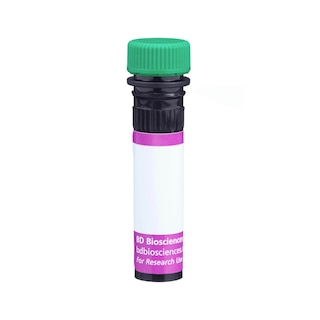
BD Horizon™ BV510 Hamster Anti-Mouse CD54

Regulatory Status Legend
Any use of products other than the permitted use without the express written authorization of Becton, Dickinson and Company is strictly prohibited.
Preparation And Storage
Product Notices
- Since applications vary, each investigator should titrate the reagent to obtain optimal results.
- Source of all serum proteins is from USDA inspected abattoirs located in the United States.
- An isotype control should be used at the same concentration as the antibody of interest.
- Please refer to www.bdbiosciences.com/us/s/resources for technical protocols.
- Caution: Sodium azide yields highly toxic hydrazoic acid under acidic conditions. Dilute azide compounds in running water before discarding to avoid accumulation of potentially explosive deposits in plumbing.
- For fluorochrome spectra and suitable instrument settings, please refer to our Multicolor Flow Cytometry web page at www.bdbiosciences.com/colors.
- Brilliant Violet™ 510 is a trademark of Sirigen.
- Although hamster immunoglobulin isotypes have not been well defined, BD Biosciences Pharmingen has grouped Armenian and Syrian hamster IgG monoclonal antibodies according to their reactivity with a panel of mouse anti-hamster IgG mAbs. A table of the hamster IgG groups, Reactivity of Mouse Anti-Hamster Ig mAbs, may be viewed at http://www.bdbiosciences.com/documents/hamster_chart_11x17.pdf.
Companion Products


The 3E2 monoclonal antibody specifically binds to CD54 (ICAM-1), a 95-kDa member of the Ig superfamily found on lymphocytes, vascular endothelium, high endothelial venules, epithelial cells, macrophages, and dendritic cells. ICAM-1 is a ligand for LFA1 (CD11a/CD18) and Mac-1 (CD11b/CD18). Its expression is upregulated upon stimulation by inflammatory mediators such as cytokines and LPS. Studies with mouse Icam1-transfected antigen-presenting cells, with CD54-blocking antibodies, and in CD54-deficient mice indicate that CD54 participates in inflammatory reactions and antigen-specific immune responses. In addition, there is evidence that CD54 is a receptor involved in MHC-non-restricted responses to weakly immunogenic tumor cells. The 3E2 antibody has been reported to block in vitro and in vivo intracellular adhesion events involved in immune responses.
The antibody was conjugated to BD Horizon™ BV510 which is part of the BD Horizon™ Brilliant Violet™ family of dyes. With an Ex Max of 405-nm and Em Max at 510-nm, BD Horizon™ BV510 can be excited by the violet laser and detected in the BD Horizon™ V500 (525/50-nm) filter set. BD Horizon™ BV510 conjugates are useful for the detection of dim markers off the violet laser.

Development References (12)
-
Gonzalo JA, Martinez C, Springer TA, Gutierrez-Ramos JC. ICAM-1 is required for T cell proliferation but not for anergy or apoptosis induced by Staphylococcus aureus enterotoxin B in vivo. Int Immunol. 1995; 7(10):1691-1698. (Clone-specific: Flow cytometry). View Reference
-
Isobe M, Yagita H, Okumura K, Ihara A. Specific acceptance of cardiac allograft after treatment with antibodies to ICAM-1 and LFA-1. Science. 1992; 255(5048):1125-1127. (Biology). View Reference
-
Kelly KJ, Williams WW Jr, Colvin RB, et al. Intercellular adhesion molecule-1-deficient mice are protected against ischemic renal injury. J Clin Invest. 1996; 97(4):1056-1063. (Biology). View Reference
-
Masten BJ, Yates JL, Pollard Koga AM, Lipscomb MF. Characterization of accessory molecules in murine lung dendritic cell function: roles for CD80, CD86, CD54, and CD40L. Am J Respir Cell Mol Biol. 1997; 16(3):335-342. (Clone-specific). View Reference
-
Nishio M, Podack ER. Rapid induction of tumor necrosis factor cytotoxicity in naive splenic T cells by simultaneous CD80 (B7.1) and CD54 (ICAM-1) co-stimulation. Eur J Immunol. 1996; 26(9):2160-2164. (Biology). View Reference
-
Nishio M, Spielman J, Lee RK, Nelson DL, Podack ER. CD80 (B7.1) and CD54 (intracellular adhesion molecule-1) induce target cell susceptibility to promiscuous cytotoxic T cell lysis. J Immunol. 1996; 157(10):4347-4353. (Biology). View Reference
-
Scheynius A, Camp RL, Pure E. Reduced contact sensitivity reactions in mice treated with monoclonal antibodies to leukocyte function-associated molecule-1 and intercellular adhesion molecule-1. J Immunol. 1993; 150(2):655-663. (Clone-specific: Flow cytometry, Inhibition, In vivo exacerbation). View Reference
-
Scheynius A, Camp RL, Pure E. Unresponsiveness to 2,4-dinitro-1-fluoro-benzene after treatment with monoclonal antibodies to leukocyte function-associated molecule-1 and intercellular adhesion molecule-1 during sensitization. J Immunol. 1996; 154(5):1804-1809. (Biology). View Reference
-
Siu G, Hedrick SM, Brian AA. Isolation of the murine intercellular adhesion molecule 1 (ICAM-1) gene. ICAM-1 enhances antigen-specific T cell activation. J Immunol. 1989; 143(11):3813-3820. (Biology). View Reference
-
Springer TA. Adhesion receptors of the immune system. Nature. 1990; 346(6283):425-434. (Clone-specific). View Reference
-
Springer TA. Traffic signals for lymphocyte recirculation and leukocyte emigration: the multistep paradigm. Cell. 1994; 76(2):301-314. (Biology). View Reference
-
Xu H, Gonzalo JA, St Pierre Y, et al. Leukocytosis and resistance to septic shock in intercellular adhesion molecule 1-deficient mice. J Exp Med. 1994; 180(1):95-109. (Clone-specific: Flow cytometry, Immunohistochemistry). View Reference
Please refer to Support Documents for Quality Certificates
Global - Refer to manufacturer's instructions for use and related User Manuals and Technical data sheets before using this products as described
Comparisons, where applicable, are made against older BD Technology, manual methods or are general performance claims. Comparisons are not made against non-BD technologies, unless otherwise noted.
For Research Use Only. Not for use in diagnostic or therapeutic procedures.