-
Reagents
- Flow Cytometry Reagents
-
Western Blotting and Molecular Reagents
- Immunoassay Reagents
-
Single-Cell Multiomics Reagents
- BD® OMICS-Guard Sample Preservation Buffer
- BD® AbSeq Assay
- BD® OMICS-One Immune Profiler Protein Panel
- BD® Single-Cell Multiplexing Kit
- BD Rhapsody™ ATAC-Seq Assays
- BD Rhapsody™ Whole Transcriptome Analysis (WTA) Amplification Kit
- BD Rhapsody™ TCR/BCR Next Multiomic Assays
- BD Rhapsody™ Targeted mRNA Kits
- BD Rhapsody™ Accessory Kits
-
Functional Assays
-
Microscopy and Imaging Reagents
-
Cell Preparation and Separation Reagents
-
- BD® OMICS-Guard Sample Preservation Buffer
- BD® AbSeq Assay
- BD® OMICS-One Immune Profiler Protein Panel
- BD® Single-Cell Multiplexing Kit
- BD Rhapsody™ ATAC-Seq Assays
- BD Rhapsody™ Whole Transcriptome Analysis (WTA) Amplification Kit
- BD Rhapsody™ TCR/BCR Next Multiomic Assays
- BD Rhapsody™ Targeted mRNA Kits
- BD Rhapsody™ Accessory Kits
- United States (English)
-
Change country/language
Old Browser
This page has been recently translated and is available in French now.
Looks like you're visiting us from {countryName}.
Would you like to stay on the current country site or be switched to your country?


.png)

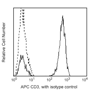
Flow cytometric analysis of C3a Receptor (C3aR) expressed on human peripheral blood leukocytes. Whole blood was stained with PE Mouse anti-Human C3a Receptor (Cat. No. 561178), APC Mouse Anti-Human CD3 (Cat. No. 555335; Left Panel) and FITC Mouse Anti-Human CD16 (Cat. No. 555406; Right Panel) antibodies. The erythrocytes were lysed with BD PharmLyse™ Lysing Buffer (Cat. No. 555899). Two-color flow cytometric dot plots showing the correlated expression patterns of CD3 or CD16 versus C3aR were derived from gated events with the forward and side light-scatter characteristics of viable peripheral blood leukocytes. Flow cytometry was performed using a BD LSR™ II Flow Cytometry System.
.png)

BD Pharmingen™ PE Mouse anti-Human C3a Receptor
.png)
Regulatory Status Legend
Any use of products other than the permitted use without the express written authorization of Becton, Dickinson and Company is strictly prohibited.
Preparation And Storage
Product Notices
- This reagent has been pre-diluted for use at the recommended Volume per Test. We typically use 1 × 10^6 cells in a 100-µl experimental sample (a test).
- Source of all serum proteins is from USDA inspected abattoirs located in the United States.
- Caution: Sodium azide yields highly toxic hydrazoic acid under acidic conditions. Dilute azide compounds in running water before discarding to avoid accumulation of potentially explosive deposits in plumbing.
- For fluorochrome spectra and suitable instrument settings, please refer to our Multicolor Flow Cytometry web page at www.bdbiosciences.com/colors.
- Please refer to http://regdocs.bd.com to access safety data sheets (SDS).
- Please refer to www.bdbiosciences.com/us/s/resources for technical protocols.
Companion Products


The hC3aRZ8 monoclonal antibody specifically binds to the human C3a Receptor (C3aR). C3aR is a seven-transmembrane glycoprotein, G-protein-coupled receptor that is the specific receptor for C3a anaphylatoxin. The C3aR consists of 482 amino acids forming a single polypeptide chain that is encoded by the C3AR1 gene located on chromosome 12 (location 12p13.31). C3aR are expressed by eosinophils, basophils, neutrophils, dendritic cells, monocytes, macrophages, endothelial cells and some T cells. C3a is a bioactive cleavage product released from Complement Component 3 (C3) during complement activation. C3a plays a role in a variety of cellular immune responses as well as being a potent pro-inflammatory agent. In response to bound C3a, this receptor stimulates cellular responses including chemotaxis, granule enzyme release and superoxide anion production and causes increased vascular permeability. In vivo, C3a production can initiate, contribute to, or exacerbate the inflammatory reactions seen in gram-negative bacterial sepsis, trauma, ARDS, ischemic heart disease, post-dialysis syndrome, and several autoimmune diseases including rheumatoid arthritis, lupus erythematosus, and acute glomerulonephritis.

Development References (8)
-
Ames RS, Li Y, Sarau HM. Molecular cloning and characterization of the human anaphylatoxin C3a receptor. J Biol Chem. 1996; 271(34):20231-20234. (Biology). View Reference
-
Crass T, Raffetseder U, Martin U. Expression cloning of the human C3a anaphylatoxin receptor (C3aR) from differentiated U-937 cells. Eur J Immunol. 1996; 26(8):1944-1950. (Biology). View Reference
-
Ember JA, Hugli TE. C3a Receptor. In: Oppenheim JJ, Feldmann M, Durum SK. editors in chief, Joost J. Oppenheim, Marc Feldman ; editors, Scott K. Durum .. et al., ed. Cytokine reference : a compendium of cytokines and other mediators of host defense. London: Academic Press; 2001:2173-2181.
-
Sacks SH. Complement fragments C3a and C5a: The salt and pepper of the immune response. Eur J Immunol. 2010; 40(3):668-670. (Biology). View Reference
-
Soruri A, Kiafard Z, Dettmer C, Riggert J, Köhl J, Zwirner J. IL-4 down-regulates anaphylatoxin receptors in monocytes and dendritic cells and impairs anaphylatoxin-induced migration in vivo.. J Immunol. 2003; 170(6):3306-14. (Immunogen). View Reference
-
Strainic MG, Liu J, Huang D, et al. Locally produced complement fragments C5a and C3a provide both costimulatory and survival signals to naive CD4+ T cells.. Immunity. 2008; 28(3):425-35. (Biology). View Reference
-
Werfel T, Kirchhoff K, Wittmann M, et al. Activated human T lymphocytes express a functional C3a receptor. J Immunol. 2000; 165(11):6599-6605. (Biology). View Reference
-
Zwirner J, Gotze O, Begemann G, Kapp A, Kirchhoff K, Werfel T. Evaluation of C3a receptor expression on human leucocytes by the use of novel monoclonal antibodies. Immunology. 1999; 97(1):166-172. (Biology). View Reference
Please refer to Support Documents for Quality Certificates
Global - Refer to manufacturer's instructions for use and related User Manuals and Technical data sheets before using this products as described
Comparisons, where applicable, are made against older BD Technology, manual methods or are general performance claims. Comparisons are not made against non-BD technologies, unless otherwise noted.
For Research Use Only. Not for use in diagnostic or therapeutic procedures.