-
Reagents
- Flow Cytometry Reagents
-
Western Blotting and Molecular Reagents
- Immunoassay Reagents
-
Single-Cell Multiomics Reagents
- BD® OMICS-Guard Sample Preservation Buffer
- BD® AbSeq Assay
- BD® OMICS-One Immune Profiler Protein Panel
- BD® Single-Cell Multiplexing Kit
- BD Rhapsody™ ATAC-Seq Assays
- BD Rhapsody™ Whole Transcriptome Analysis (WTA) Amplification Kit
- BD Rhapsody™ TCR/BCR Next Multiomic Assays
- BD Rhapsody™ Targeted mRNA Kits
- BD Rhapsody™ Accessory Kits
-
Functional Assays
-
Microscopy and Imaging Reagents
-
Cell Preparation and Separation Reagents
-
- BD® OMICS-Guard Sample Preservation Buffer
- BD® AbSeq Assay
- BD® OMICS-One Immune Profiler Protein Panel
- BD® Single-Cell Multiplexing Kit
- BD Rhapsody™ ATAC-Seq Assays
- BD Rhapsody™ Whole Transcriptome Analysis (WTA) Amplification Kit
- BD Rhapsody™ TCR/BCR Next Multiomic Assays
- BD Rhapsody™ Targeted mRNA Kits
- BD Rhapsody™ Accessory Kits
- United States (English)
-
Change country/language
Old Browser
This page has been recently translated and is available in French now.
Looks like you're visiting us from {countryName}.
Would you like to stay on the current country site or be switched to your country?


.png)

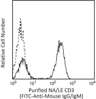
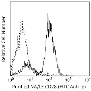
Analysis of SHP2 (pY542) in activated human lymphocytes. Peripheral blood mononuclear cells (PBMC, left panel) were either stimulated by cross-linking of CD3 and CD28 with NA/LE Mouse anti-Human CD3 mAb UCHT1 (Cat. No. 555329) and NA/LE Mouse anti-Human CD28 mAb CD28.2 (Cat. No. 555725) on ice for 15 minutes followed by Purified Goat anti-Mouse Ig (Cat. No. 553998) on ice for 15 minutes, then allowed to undergo phosphorylation at 37°C for 1-2 minutes (shaded histogram) or unstimulated (open histogram). The PBMC were then fixed with BD Cytofix™ Fixation Buffer (Cat. No. 554655) for 10 minutes at 37ºC, permeabilized with BD Phosflow™ Perm Buffer III (Cat. No. 558050) on ice for at least 30 minutes, blocked with normal mouse immunoglobulin, and then stained with PE Mouse anti-Human SHP2 (Y542, Cat. No. 560389). For data analysis, lymphocytes were selected by their scatter profile. Flow cytometry was performed on a BD FACSCanto™ II flow cytometry system. The specificity of mAb L99-921 was confirmed by western blot analysis using unconjugated antibody on lysates from mouse and human cells. Lysates from NIH-3T3 cells (middle panel) that had been treated with 200 nM PDGF (Cat. No. 354051) for 30 minutes (right blot) or untreated (left blot) were probed with purified mouse anti-SHP2 (pY542) monoclonal antibody at concentrations of 1.0, 0.25 and 0.063 μg/ml (Lanes 1, 2, and 3, respectively). SHP2 (pY542) is identified as a band of 65-72 kDa in the treated cells. Lysates from PBMC (right panel) that were untreated (lane 1) or stimulated by cross-linking of CD3 and CD28 (lane 2) were probed with 0.5 μg/ml purified mouse anti-SHP2 (pY542) monoclonal antibody. SHP2 (pY542) is identified as a band of 65-72 kDa, with increased intensity in the stimulated cells.

.png)

BD™ Phosflow PE Mouse anti-SHP2 (pY542)

BD™ Phosflow PE Mouse anti-SHP2 (pY542)
.png)
Regulatory Status Legend
Any use of products other than the permitted use without the express written authorization of Becton, Dickinson and Company is strictly prohibited.
Preparation And Storage
Recommended Assay Procedures
This antibody conjugate is suitable for intracellular staining of cell lines and peripheral blood mononuclear cells using BD Cytofix™ Fixation Buffer. Any of the three BD Phosflow™ permeabilization buffers may be used.
Product Notices
- This reagent has been pre-diluted for use at the recommended Volume per Test. We typically use 1 × 10^6 cells in a 100-µl experimental sample (a test).
- Source of all serum proteins is from USDA inspected abattoirs located in the United States.
- Caution: Sodium azide yields highly toxic hydrazoic acid under acidic conditions. Dilute azide compounds in running water before discarding to avoid accumulation of potentially explosive deposits in plumbing.
- Ficoll-Paque is a trademark of Amersham Biosciences Limited.
- For fluorochrome spectra and suitable instrument settings, please refer to our Multicolor Flow Cytometry web page at www.bdbiosciences.com/colors.
- Please refer to www.bdbiosciences.com/us/s/resources for technical protocols.
Companion Products


SHP2 is a member of the cytosolic class of protein-tyrosine phosphatases (PTPs). SHP2 reportedly contains two SH2 domains, both of which are N-terminal to the PTP catalytic domain. SH2 PTPs are believed to work in conjunction with protein-tyrosine kinases to maintain intracellular protein phosphotyrosine homeostasis and cell cycle progression. The expression of SHP2 has been reported to be highest in brain, heart, and kidney. SHP2 is phosphorylated at two C-terminal tyrosine residues: Y542 and Y580. Phosphorylation of Y542 creates an interaction in the N-terminal SH2 domain and appears to relieve basal inhibition of the PTP, while phosphorylation at Y580 creates an interaction at the C-terminal that stimulates PTP activity.
The L99-921 monoclonal antibody recognizes the phosphorylated Y542 of SHP2 (PTP2C isoform 2). The homologous phosphorylation site in the PTP2Ci splice variant (isoform 1) is Y546. The specificity of this antibody was validated by confirming, using western blot analysis, that RNA-mediated interference (RNAi) of the specific protein was able to down-regulate the expression of SHP2 (pY542).

Development References (6)
-
Lu W, Gong D, Bar-Sagi D, Cole PA. Site-specific incorporation of a phosphotyrosine mimetic reveals a role for tyrosine phosphorylation of SHP-2 in cell signaling. Mol Cell. 2001; 8:759-769. (Biology). View Reference
-
Ahmad S, Banville D, Zhao Z, Fischer EH, Shen SH. A widely expressed human protein-tyrosine phosphatase containing src homology 2 domains. Proc Natl Acad Sci U S A. 1993; 90(6):2197-2201. (Biology). View Reference
-
Bennett AM, Tang TL, Sugimoto S, Walsh CT, Neel BG. Protein-tyrosine-phosphatase SHPTP2 couples platelet-derived growth factor receptor beta to Ras. Proc Natl Acad Sci U S A. 1994; 91(15):7335-7339. (Biology). View Reference
-
Fornasa G, Groyer E, Clement M, et al. TCR stimulation drives cleavage and shedding of the ITIM receptor CD31. J Immunol. 2010; 184(10):5485-5492. (Clone-specific: Flow cytometry). View Reference
-
MacGillivray M, Herrera-Abreu MT, Chow CW et al. The protein tyrosine phosphatase SHP-2 regulates interleukin-1-induced ERK activation in fibroblasts. J Biol Chem. 2003; 278(29):27190-27198. (Biology). View Reference
-
Marie-Cardine A, Kirchgessner H, Bruyns E et al. SHP2-interacting transmembrane adaptor protein (SIT), a novel disulfide-linked dimer regulating human T cell activation. J Exp Med. 1999; 189(8):1181-1194. (Biology). View Reference
Please refer to Support Documents for Quality Certificates
Global - Refer to manufacturer's instructions for use and related User Manuals and Technical data sheets before using this products as described
Comparisons, where applicable, are made against older BD Technology, manual methods or are general performance claims. Comparisons are not made against non-BD technologies, unless otherwise noted.
For Research Use Only. Not for use in diagnostic or therapeutic procedures.