-
Reagents
- Flow Cytometry Reagents
-
Western Blotting and Molecular Reagents
- Immunoassay Reagents
-
Single-Cell Multiomics Reagents
- BD® OMICS-Guard Sample Preservation Buffer
- BD® AbSeq Assay
- BD® OMICS-One Immune Profiler Protein Panel
- BD® Single-Cell Multiplexing Kit
- BD Rhapsody™ ATAC-Seq Assays
- BD Rhapsody™ Whole Transcriptome Analysis (WTA) Amplification Kit
- BD Rhapsody™ TCR/BCR Next Multiomic Assays
- BD Rhapsody™ Targeted mRNA Kits
- BD Rhapsody™ Accessory Kits
-
Functional Assays
-
Microscopy and Imaging Reagents
-
Cell Preparation and Separation Reagents
-
- BD® OMICS-Guard Sample Preservation Buffer
- BD® AbSeq Assay
- BD® OMICS-One Immune Profiler Protein Panel
- BD® Single-Cell Multiplexing Kit
- BD Rhapsody™ ATAC-Seq Assays
- BD Rhapsody™ Whole Transcriptome Analysis (WTA) Amplification Kit
- BD Rhapsody™ TCR/BCR Next Multiomic Assays
- BD Rhapsody™ Targeted mRNA Kits
- BD Rhapsody™ Accessory Kits
- United States (English)
-
Change country/language
Old Browser
This page has been recently translated and is available in French now.
Looks like you're visiting us from {countryName}.
Would you like to stay on the current country site or be switched to your country?


.png)

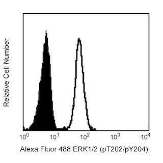
Analysis of Lck (pY505) in activated human T leukemia cells. Jurkat cells (ATCC TIB-152) were serum starved overnight and then either stimulated with 5 mM hydrogen peroxide for 15 minutes (shaded histogram) or unstimulated (open histogram). The cells were fixed with pre-warmed BD Cytofix™ buffer (Cat. No. 554655) for 10 minutes, then permeabilized (BD PhosFlow™ Perm Buffer III, Cat. No. 558050) on ice for at least 30 minutes, and then stained with PE anti-Lck (pY505). Flow cytometry was performed on a BD FACSCalibur™ flow cytometry system.
.png)

BD™ Phosflow PE Mouse Anti-Human Lck (pY505)
.png)
Regulatory Status Legend
Any use of products other than the permitted use without the express written authorization of Becton, Dickinson and Company is strictly prohibited.
Preparation And Storage
Recommended Assay Procedures
This antibody conjugate is suitable for intracellular staining of human whole blood (using BD Phosflow™ Lyse/Fix Buffer) and peripheral blood mononuclear cells and cell lines (using BD Cytofix™ Fixation Buffer or BD Phosflow™ Fix Buffer I). Any of the three BD Phosflow™ permeabilization buffers may be used.
Product Notices
- This reagent has been pre-diluted for use at the recommended Volume per Test. We typically use 1 × 10^6 cells in a 100-µl experimental sample (a test).
- Please refer to www.bdbiosciences.com/us/s/resources for technical protocols.
- For fluorochrome spectra and suitable instrument settings, please refer to our Multicolor Flow Cytometry web page at www.bdbiosciences.com/colors.
- Caution: Sodium azide yields highly toxic hydrazoic acid under acidic conditions. Dilute azide compounds in running water before discarding to avoid accumulation of potentially explosive deposits in plumbing.
- Source of all serum proteins is from USDA inspected abattoirs located in the United States.
- Species cross-reactivity detected in product development may not have been confirmed on every format and/or application.
- Human donor specific background has been observed in relation to the presence of anti-polyethylene glycol (PEG) antibodies, developed as a result of certain vaccines containing PEG, including some COVID-19 vaccines. We recommend use of BD Horizon Brilliant™ Stain Buffer in your experiments to help mitigate potential background. For more information visit https://www.bdbiosciences.com/en-us/support/product-notices.
Companion Products


Lck is a member of the Src family of cytoplasmic protein-tyrosine kinases (PTKs) that is normally expressed exclusively in lymphoid cells, primarily T lymphocytes and NK cells. Members of this family have several common features: 1) unique N-terminal domains, 2) attachment to cellular membranes through a myristylated N-terminus, and 3) homologous SH2, SH3, and catalytic domains. The unique N-terminal domain of Lck interacts with the cytoplasmic tails of the CD4 and CD8 cell-surface glycoproteins of T lymphocytes, which recognize antigen presenting cells via their surface MHC class II and class I molecules, respectively. The catalytic activity of Lck is regulated by both kinases and phosphatases that control the phosphorylation states of two tyrosine residues that have opposing effects. Repression of Lck's catalytic activity occurs via phosphorylation at tyrosine 505 (Y505), located near the carboxy terminus. Phosphorylation of this tyrosine site is mediated by the Csk family of PTKs, and its dephosphorylation is mediated by the protein tyrosine phosphatase, CD45. When Lck is phosphorylated at this site, it assumes a folded tertiary structure which is enzymatically inactive. When CD45 dephosphorylates it at Y505, Lck is able to autophosphorylate its Y394, which leads to conformational changes in the catalytic domain that induce kinase activity. However, it has been observed that the inhibitory effect of the phosphorylated Y505 can be overcome by direct engagement of Lck's SH3 domain and that both Y394 and Y505 are phosphorylated together in cells activated by hydrogen peroxide. Activated Lck phosphorylates the ITAMs (Immunoreceptor-based Tyrosine Activation Motifs) of the T cell receptor (TCR) and thus is critical for activation and development of T lymphocytes. The interactions of Lck, Csk, CD45, CD4 or CD8, and TCR are only a small part of a complex immunoregulatory cascade that involves additional substrates for Csk and CD45, other enzymes, adhesion molecules, adaptor proteins, and specialized membrane microdomains.
The 4/LCK-Y505 monoclonal antibody recognizes the phosphorylated Y505 of the catalytic domain of Lck. The Alexa Fluor® 488- conjugated format has been evaluated by flow using a human model system. However, the unconjugated form of this antibody (Cat. No. 612390) has been shown to react with human, mouse, and rat in western blot. A phosphorylated peptide corresponding to residues around Tyrosine-505 from human Lck was used as the immunogen.

Development References (5)
-
Hardwick JS, Sefton BM. The activated form of the Lck tyrosine protein kinase in cells exposed to hydrogen peroxide is phosphorylated at both Tyr-394 and Tyr-505. J Biol Chem. 1997; 272:25429-25432. (Biology).
-
Holdorf AD, Lee K-H, Burack WR, Allen PM, Shaw AS. Regulation of Lck activity by CD4 and CD28 in the immunological synapse. Nat Immunol. 2002; 3(3):259-264. (Biology).
-
Johnson KG, Bromley SK, Dustin ML, Thomas ML. A supramolecular basis for CD45 tyrosine phosphatase regulation in sustained T cell activation. Proc Natl Acad Sci U S A. 2000; 97:10138-10143. (Biology).
-
Lee-Fruman KK, Collins TL, Burakoff SJ. Role of the Lck Src homology 2 and 3 domains in protein tyrosine phosphorylation. J Biol Chem. 1996; 271:25003-25010. (Biology).
-
Veillette A, Latour S, Davidson D. Negative regulation of immunoreceptor signaling. Annu Rev Immunol. 2002; 20:669-707. (Biology).
Please refer to Support Documents for Quality Certificates
Global - Refer to manufacturer's instructions for use and related User Manuals and Technical data sheets before using this products as described
Comparisons, where applicable, are made against older BD Technology, manual methods or are general performance claims. Comparisons are not made against non-BD technologies, unless otherwise noted.
For Research Use Only. Not for use in diagnostic or therapeutic procedures.