-
Reagents
- Flow Cytometry Reagents
-
Western Blotting and Molecular Reagents
- Immunoassay Reagents
-
Single-Cell Multiomics Reagents
- BD® OMICS-Guard Sample Preservation Buffer
- BD® AbSeq Assay
- BD® OMICS-One Immune Profiler Protein Panel
- BD® Single-Cell Multiplexing Kit
- BD Rhapsody™ ATAC-Seq Assays
- BD Rhapsody™ Whole Transcriptome Analysis (WTA) Amplification Kit
- BD Rhapsody™ TCR/BCR Next Multiomic Assays
- BD Rhapsody™ Targeted mRNA Kits
- BD Rhapsody™ Accessory Kits
-
Functional Assays
-
Microscopy and Imaging Reagents
-
Cell Preparation and Separation Reagents
-
- BD® OMICS-Guard Sample Preservation Buffer
- BD® AbSeq Assay
- BD® OMICS-One Immune Profiler Protein Panel
- BD® Single-Cell Multiplexing Kit
- BD Rhapsody™ ATAC-Seq Assays
- BD Rhapsody™ Whole Transcriptome Analysis (WTA) Amplification Kit
- BD Rhapsody™ TCR/BCR Next Multiomic Assays
- BD Rhapsody™ Targeted mRNA Kits
- BD Rhapsody™ Accessory Kits
- United States (English)
-
Change country/language
Old Browser
This page has been recently translated and is available in French now.
Looks like you're visiting us from {countryName}.
Would you like to stay on the current country site or be switched to your country?


Western blot analysis of calcineurin. Lysate from A-431 cells was probed with anti-calcineurin (clone G182-1847) at concentrations of 1.0 (lane 1), 0.2 (lane 2), and 0.04 µg/ml (lane 3). The 61 kDa A subunit of calcineurin is identified.


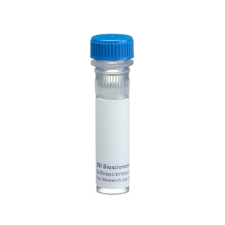
BD Pharmingen™ Purified Mouse Anti- Calcineurin

Regulatory Status Legend
Any use of products other than the permitted use without the express written authorization of Becton, Dickinson and Company is strictly prohibited.
Preparation And Storage
Recommended Assay Procedures
Applications include immunoprecipitation (1-2 µg/ml) and western blot analysis (1-2 µg/ml). Jurkat human T cells (ATCC TIB-152)2, HeLa human carcinoma cells (ATCC CCL-2), and A-431 human epidermal carcinoma cells (ATCC CRL-1555) are suggested as positive controls. A431 cell lysate (Cat. No. 611447) and HeLa cell lysate (Cat. No. 611449) are also available as ready-to-use positive western blot control.
Product Notices
- Since applications vary, each investigator should titrate the reagent to obtain optimal results.
- Please refer to www.bdbiosciences.com/us/s/resources for technical protocols.
- Caution: Sodium azide yields highly toxic hydrazoic acid under acidic conditions. Dilute azide compounds in running water before discarding to avoid accumulation of potentially explosive deposits in plumbing.
Companion Products


Calcineurin is a Ca2+- and calmodulin-dependent serine/threonine phosphatase which consists of two subunits. The A subunit (~61 kDa) is catalytic and has calmodulinbinding activity; the B subunit (~19 kDa), which is myristylated at its N-terminus, has Ca[2+]-binding activity. Calcineurin is expressed ubiquitously in eukaryotic cells, including yeast. The protein is highly expressed in mammalian brain, and has been implicated in a number of calcium-dependent pathways in the nervous system, including regulation of neurotransmitter release. Calcineurin also plays a key role in T cell receptor-mediated signal transduction pathways. Calcineurin dephosphorylates components of NF-AT (Nuclear Factor of Activated T cells), allowing NF-AT to translocate to the nucleus where it participates with AP-1 in activation of multiple cytokine and surface receptor genes. Inhibition of calcineurin by immunosuppressive agents such as cyclosporin A and FK506 correlates with the suppression of early T cell activation events. These immunosuppressive agents bind to specific intracellular proteins, called immunophilins, to form complexes that inhibit calcineurin's phosphatase activity. In the presence of these complexes, NF-AT remains phosphorylated and thus cannot translocate to the nucleus. A novel protein, Cain (calcineurin inhibitor), has been described which displays a similar pattern of neuronal and brain expression and has been shown to bind and inhibit calcineurin activity in vivo.
G182-1847 recognizes the α and β isoforms of the A subunit of calcineurin (~61 kDa). It reacts with an epitope in the autoinhibitory domain. It does not recognize the B subunit of calcineurin. A recombinant tag-fusion protein containing amino acids 349-505 of the human β1 isoform of calcineurin A subunit was used as immunogen.
Development References (3)
-
Fruman DA, Klee CB, Bierer BE, Burakoff SJ. Calcineurin phosphatase activity in T lymphocytes is inhibited by FK 506 and cyclosporin A. Proc Natl Acad Sci U S A. 1992; 89(9):3686-3690. (Biology). View Reference
-
Husi H, Luyten MA, Zurini MG. Mapping of the immunophilin-immunosuppressant site of interaction on calcineurin. J Biol Chem. 1994; 269(19):14199-14204. (Biology). View Reference
-
Lai MM, Burnett PE, Wolosker H, Blackshaw S, Snyder SH. Cain, a novel physiologic protein inhibitor of calcineurin. J Biol Chem. 1998; 273(29):18325-18331. (Biology). View Reference
Please refer to Support Documents for Quality Certificates
Global - Refer to manufacturer's instructions for use and related User Manuals and Technical data sheets before using this products as described
Comparisons, where applicable, are made against older BD Technology, manual methods or are general performance claims. Comparisons are not made against non-BD technologies, unless otherwise noted.
For Research Use Only. Not for use in diagnostic or therapeutic procedures.